Biology · Blood, Blood Vessels and the Heart
Knowledge organiser
The whole chapter on two pages. Print it on A4 — they are already landscape — or cover a box and see how much of it you can say from memory.
Biology · Edexcel Paper 2
Blood, Blood Vessels and the Heart
The Structure and Functions of Blood
Every part of the blood has a structure that suits its job: red blood cells carry oxygen, white blood cells defend, platelets clot and plasma transports.
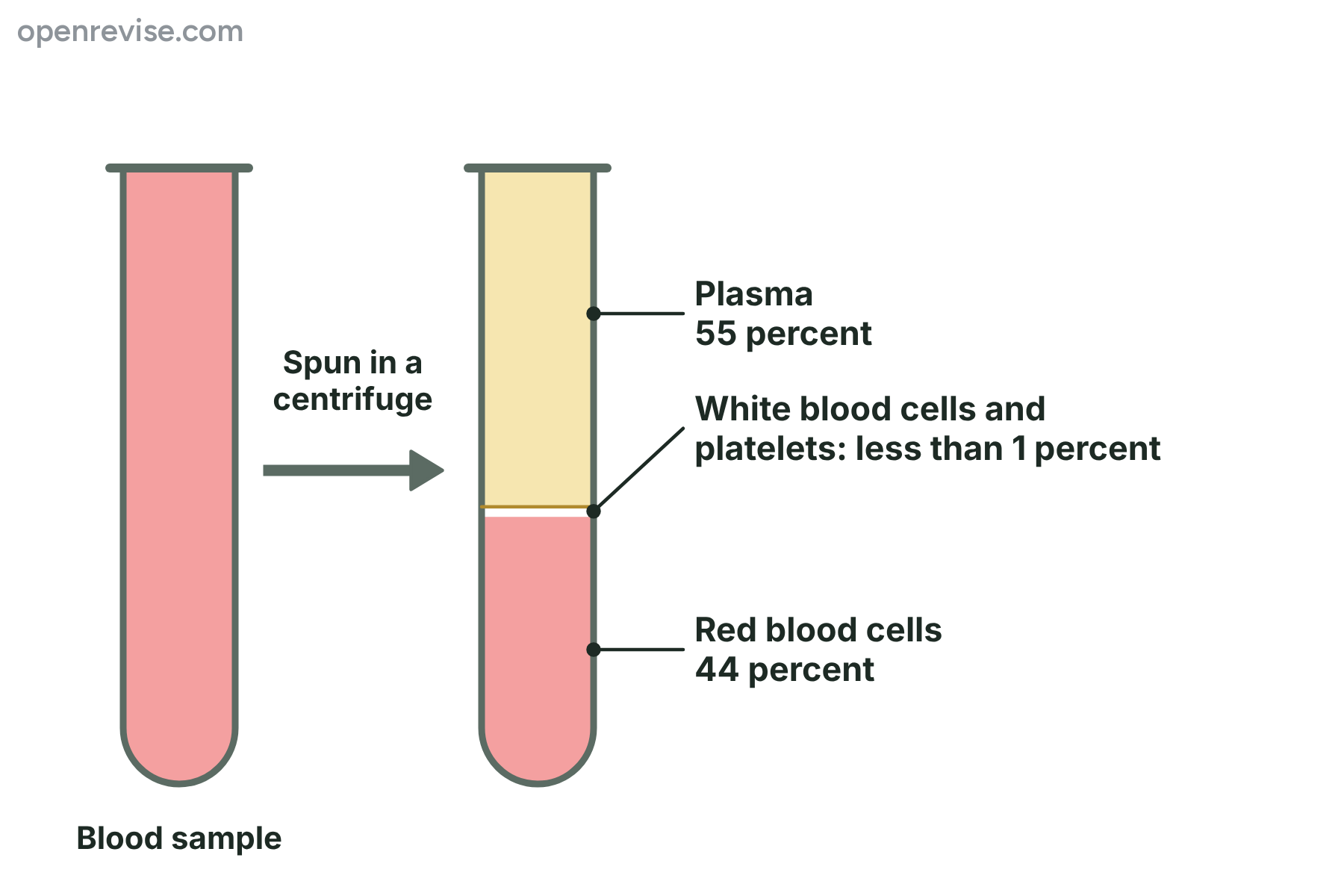
- Plasma The pale yellow liquid part of the blood, which carries dissolved substances, hormones, heat and blood cells.
- Red blood cell A blood cell with no nucleus that is full of haemoglobin and carries oxygen (an erythrocyte).
- Haemoglobin The red protein in red blood cells that contains iron and combines with oxygen.
Red blood cells and white blood cells
- Red blood cells No nucleus; Packed with haemoglobin; A biconcave disc about 7 micrometres across
- White blood cells A nucleus, which may have lobes; No haemoglobin; Round or irregular, and larger than red cells
How a blood clot forms
- The vessel is cut Blood leaks out and the damaged wall is exposed.
- Platelets collect Platelets stick to the damaged area and release chemicals.
- A mesh forms The chemicals change the soluble protein fibrinogen into insoluble threads of fibrin.
- The mesh traps cells Red blood cells and platelets are trapped in the mesh, which forms a clot.
Arteries, Veins and Capillaries
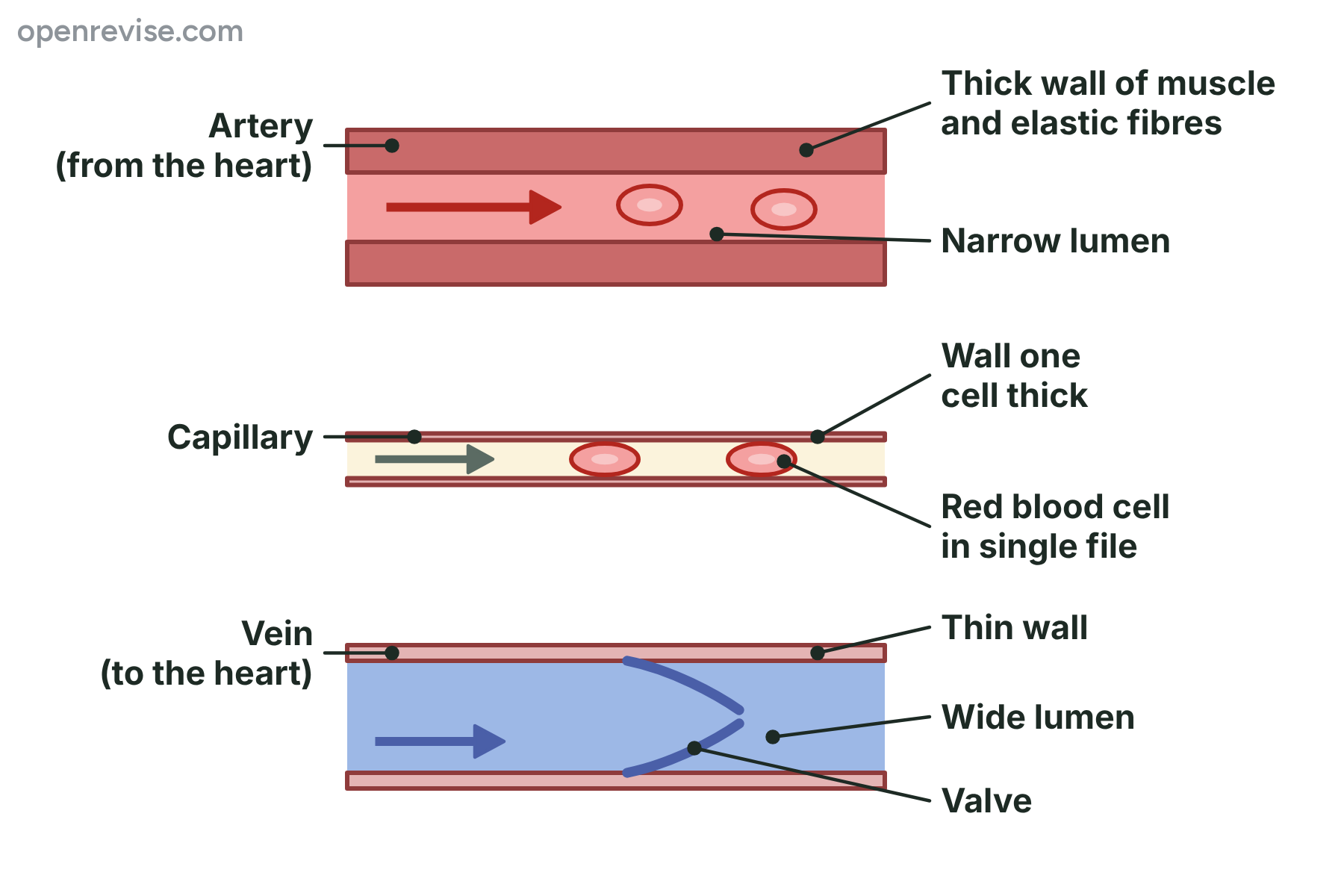
Arteries are strong, veins are wide with valves, and capillaries are thin and tiny, because the structure of each vessel suits its job.
- Artery A blood vessel that carries blood away from the heart at high pressure.
- Vein A blood vessel that carries blood towards the heart at low pressure.
- Capillary A tiny blood vessel with a wall one cell thick, where substances are exchanged with the cells.
Arteries and veins
- Arteries Carry blood away from the heart; High pressure; Thick wall with muscle and elastic fibres
- Veins Carry blood towards the heart; Low pressure; Thin wall with little muscle
The route of blood around the body
- Artery The heart pumps blood at high pressure into the arteries.
- Smaller arteries The arteries branch into smaller vessels that take the blood to each organ.
- Capillaries The smallest vessels run between the cells.
- Small veins The capillaries join up to form small veins, and the pressure is now low.
Biology · Edexcel Paper 2
Blood, Blood Vessels and the Heart continued
The Heart and Double Circulation
The heart is two pumps in one, and a double circulation sends blood to the lungs and then back to the heart so that it can be pumped to…
- Atrium An upper chamber of the heart that receives blood from the veins (plural atria).
- Ventricle A lower chamber of the heart with a thick muscular wall that pumps blood into the arteries.
- Septum The wall of muscle that separates the left and right sides of the heart.
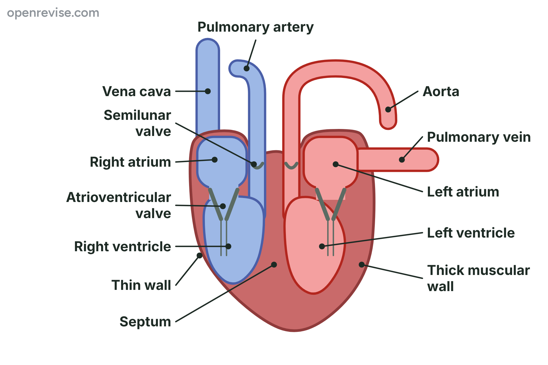
The right side and the left side of the heart
- Right side Receives deoxygenated blood from the body through the vena cava; Pumps the blood to the lungs through the pulmonary artery; The right ventricle has a thinner wall
- Left side Receives oxygenated blood from the lungs through the pulmonary vein; Pumps the blood to the body through the aorta; The left ventricle has a much thicker wall
The route of the blood
- Vena cava Deoxygenated blood from the body flows in through the vena cava.
- Right atrium to right ventricle The right atrium contracts, and the blood passes through the…
- To the lungs The right ventricle contracts and forces the blood through the…
- Gas exchange In the lungs the blood loses carbon dioxide and picks up oxygen.
Heart Rate, Stroke Volume and Cardiac Output
\text{cardiac output} = \text{stroke volume} × \text{heart rate}
- Heart rate The number of times the heart beats in one minute, measured in beats per minute.
- Stroke volume The volume of blood pumped out of a ventricle in one heartbeat.
- Cardiac output The volume of blood pumped out of the heart in one minute, equal to stroke volume times heart rate.
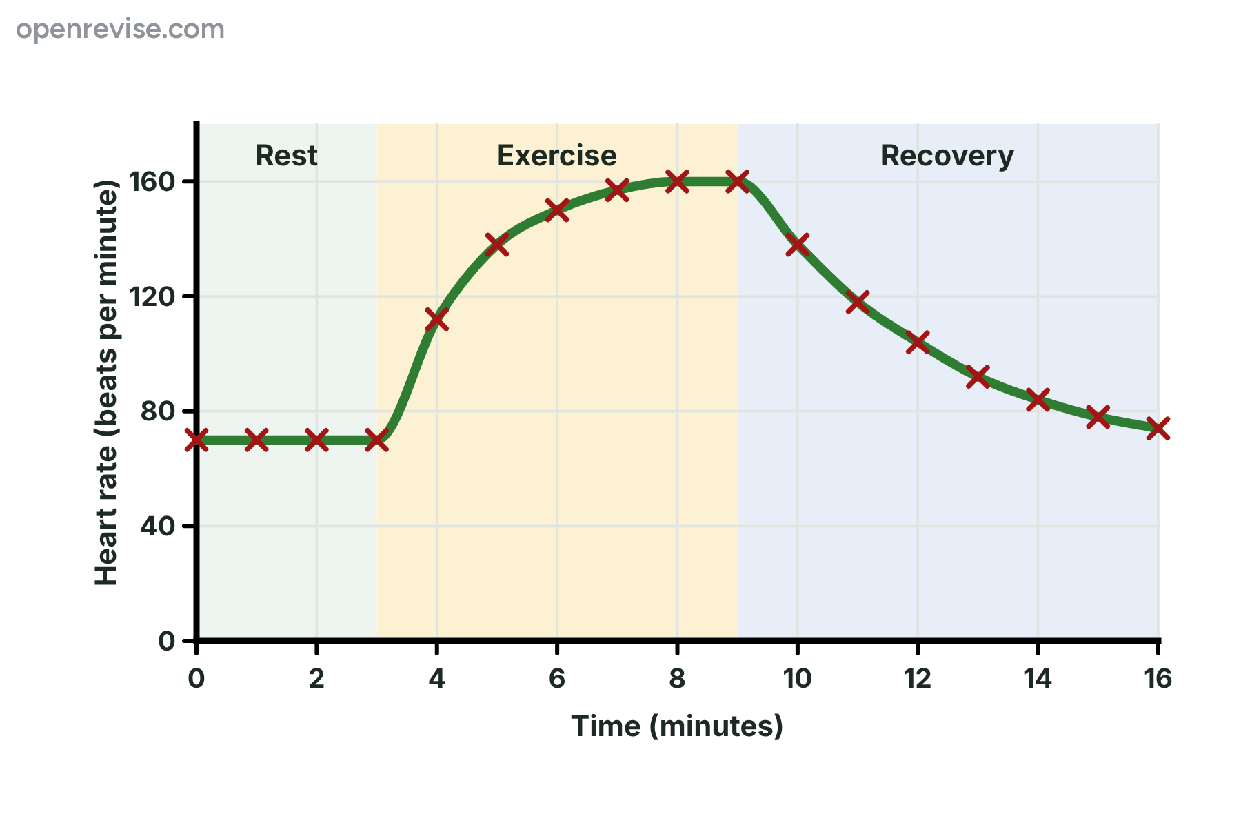
Investigating the effect of exercise on heart rate
- Rest The volunteer sits quietly for five minutes.
- Exercise The volunteer steps up and down on a step, or jogs on the spot.
- Measure straight away Immediately find the pulse again, counting for 15 seconds and multiplying by 4.
- Recovery Measure the pulse every minute until it returns to the resting value.