Biology · Cell Structures
Knowledge organiser
The whole chapter on one page. Print it on A4 — it is already landscape — or cover a box and see how much of it you can say from memory.
Biology · OCR Paper 1
Cell Structures
Eukaryotic and Prokaryotic Cells
Bacteria do have DNA. It is just not shut inside a nucleus.
- Eukaryotic cell A cell whose genetic material is enclosed in a nucleus, such as an animal or plant cell.
- Prokaryotic cell A cell with no nucleus, whose genetic material lies free in the cytoplasm, such as a bacterium.
- Nucleus The part of a eukaryotic cell that contains the genetic material and controls the cell's activities.
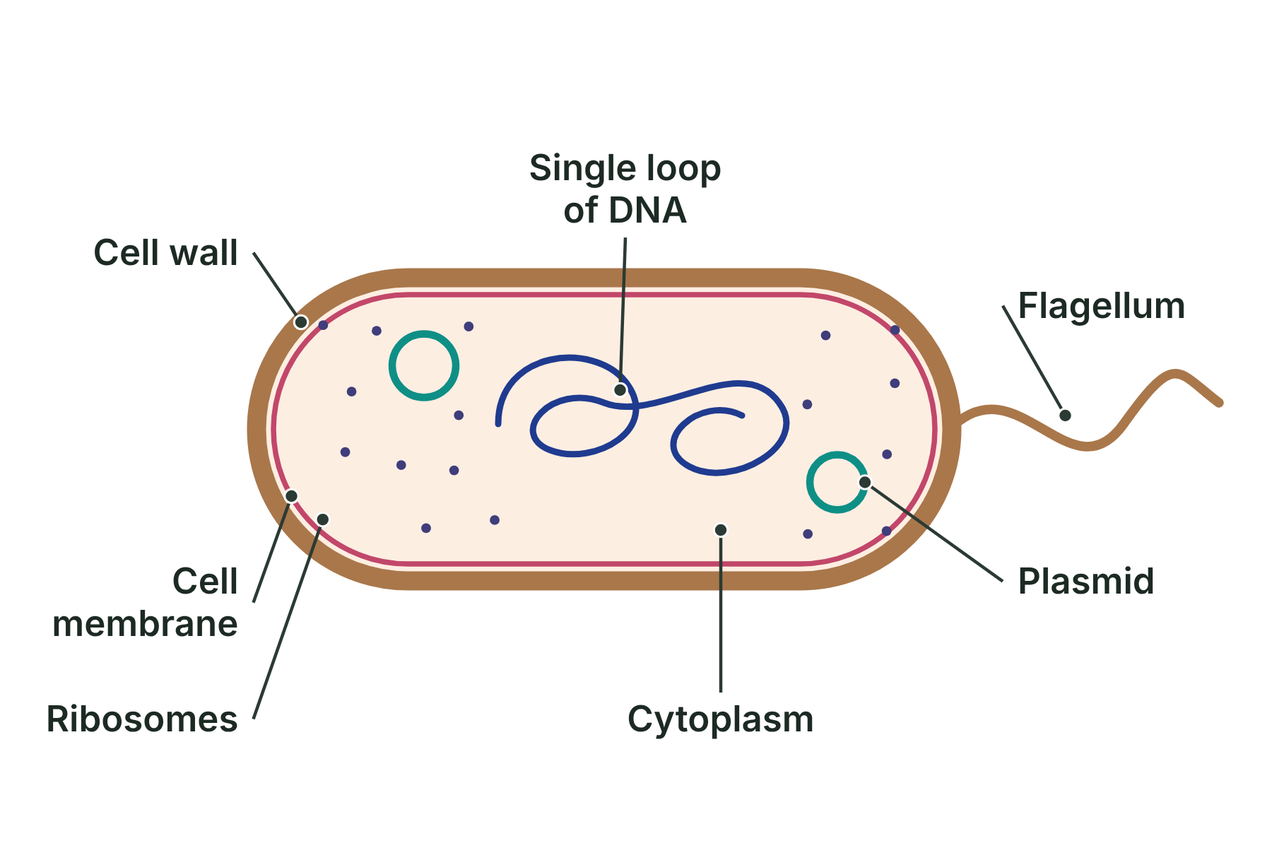
Eukaryotic and prokaryotic cells compared
- Eukaryotic cell (animal or plant) Genetic material is enclosed in a nucleus; Larger, often 10 to 100 µm across; Has a cell membrane, cytoplasm, mitochondria and ribosomes
- Prokaryotic cell (bacterium) No nucleus: a single loop of DNA lies in the cytoplasm; Much smaller, often 1 to 5 µm long; Has a cell membrane, cytoplasm and ribosomes, with a cell wall outside
Animal and Plant Cells
Structure always fits function: a cell has more of whatever its job needs most.
- Sub-cellular structure A structure inside a cell, such as the nucleus or a mitochondrion.
- Mitochondria Structures where aerobic respiration takes place, releasing energy for the cell.
- Ribosome A tiny structure where proteins are made.
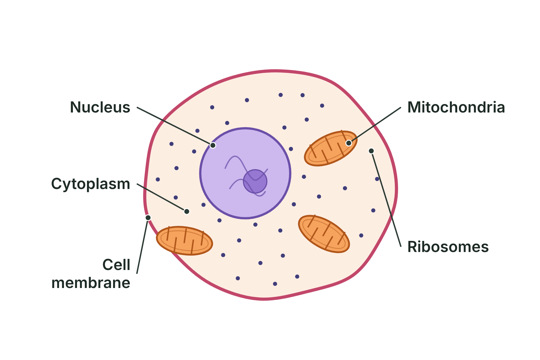
Animal cells and plant cells compared
- Animal cell Nucleus, cytoplasm, cell membrane, mitochondria, ribosomes; No cell wall, so the shape can change; No chloroplasts, so it cannot photosynthesise
- Plant cell Has all of those structures too; Cellulose cell wall gives a fixed shape; Chloroplasts in the green parts, for photosynthesis
Microscopy and Magnification
Magnification makes an image bigger. Resolution makes it clearer.
- Magnification How many times bigger an image is than the real object.
- Resolution The ability to see two points that are close together as separate. Also called resolving power.
- Light microscope A microscope that uses light and glass lenses to magnify a specimen.
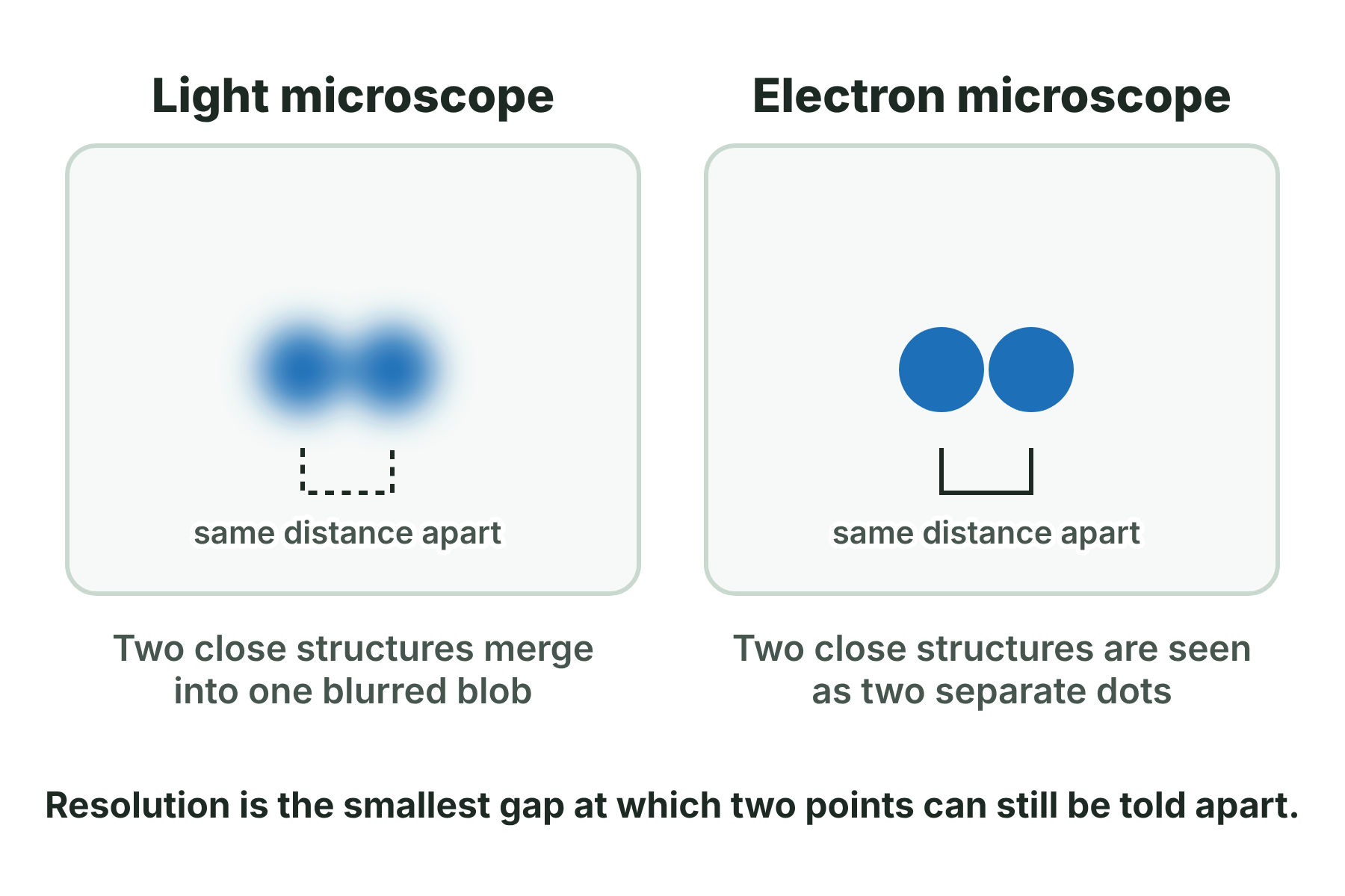
Light microscope and electron microscope compared
- Light microscope Magnifies up to about ×1500; Resolution about 0.2 µm (200 nm); Specimen can be alive, and colour stains can be used
- Electron microscope Magnifies up to about ×2 000 000; Resolution about 0.2 nm, about 1000 times better; Specimen is dead and the image is black and white
Using a light microscope (core practical)
- Prepare the slide Put a thin piece of tissue, such as onion skin, on a slide, add…
- Clip it on the stage Place the slide on the stage and secure it with the clips.
- Choose the lowest power Turn the nosepiece to the ×4 objective lens.
- Focus Looking from the side, use the coarse focus wheel to raise the…