Flashcards · Biology
The Circulatory System
-
What is the heart?
An organ that pumps blood around the body in a double circulatory system.
-
Which chamber receives blood from the vena cava?
The right atrium.
-
Which chamber pumps blood to the lungs?
The right ventricle.
-
Which chamber pumps blood to the body?
The left ventricle.
-
Why is the wall of the left ventricle thicker than the right?
It has to pump blood around the whole body, so it needs more force.
-
What does the aorta carry?
Oxygenated blood from the left ventricle to the body.
-
What does the pulmonary artery carry?
Deoxygenated blood from the right ventricle to the lungs.
-
What does the pulmonary vein carry?
Oxygenated blood from the lungs to the left atrium.
-
What do the coronary arteries do?
They supply the heart muscle with oxygenated blood.
-
Why is it called a double circulatory system?
Blood goes through the heart twice on each complete trip round the body.
-
Why is blood returned to the heart after the lungs?
It loses pressure in the lung capillaries, so the heart gives it a new push to the body.
-
Where is the natural pacemaker?
In a group of cells in the right atrium.
-
What does an artificial pacemaker do?
It sends electrical pulses to the heart to correct an irregular heartbeat.
-
How do you work out a heart rate from a count in 15 seconds?
Multiply the count by 4 to get beats per minute.
-
Do you need to know the names of the heart valves?
No. They are not required.
-
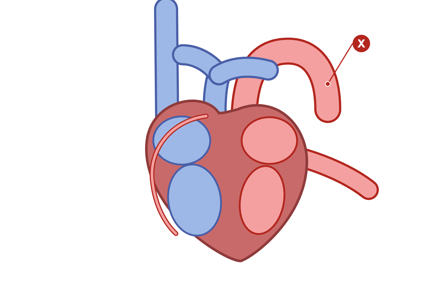 Name the vessel marked X and say what it carries.
 The aorta. It carries oxygenated blood from the left ventricle to the body.
-
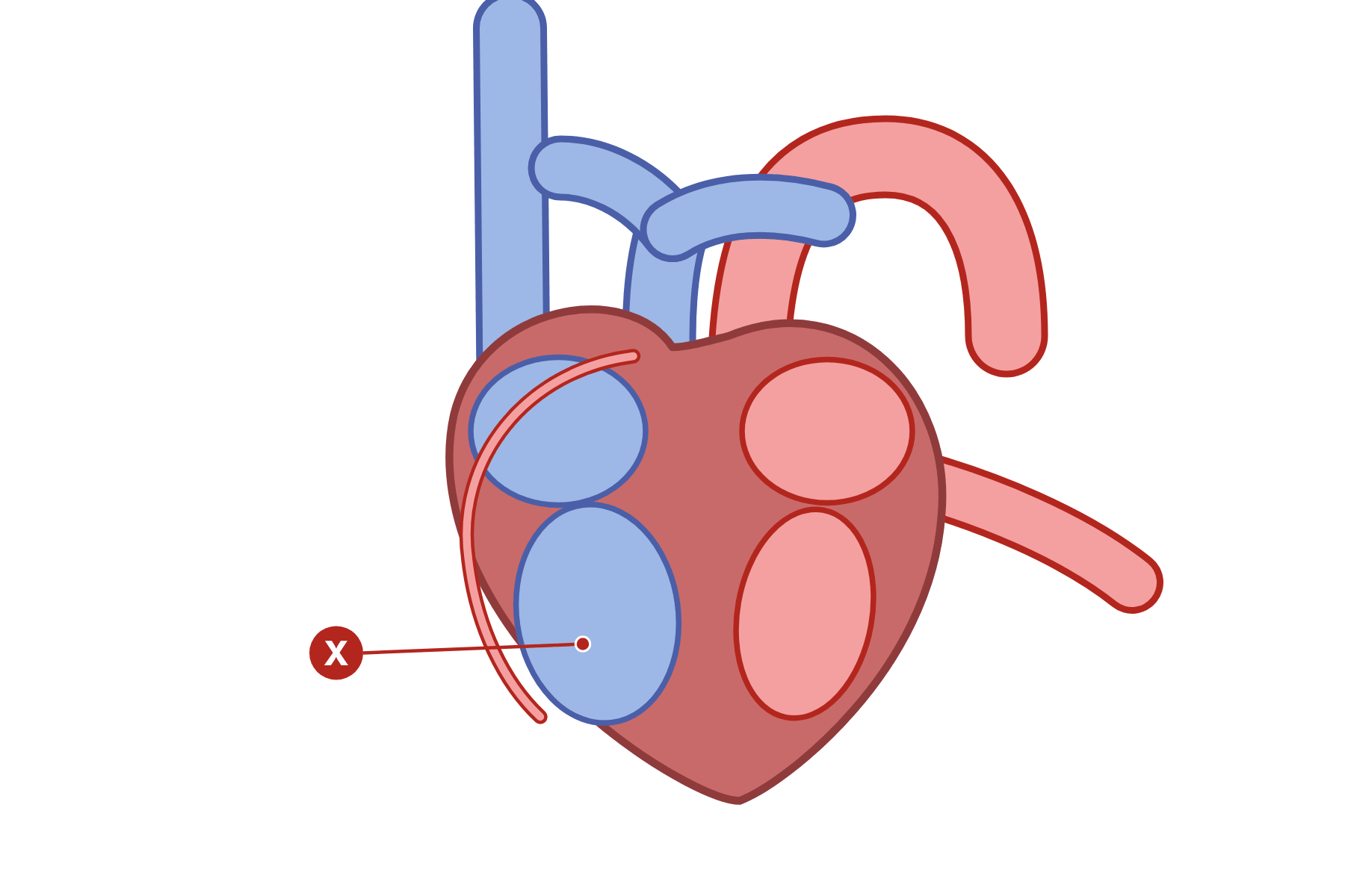 Name the chamber marked X and say where it pumps blood.
 The right ventricle. It pumps deoxygenated blood to the lungs along the pulmonary artery.
-
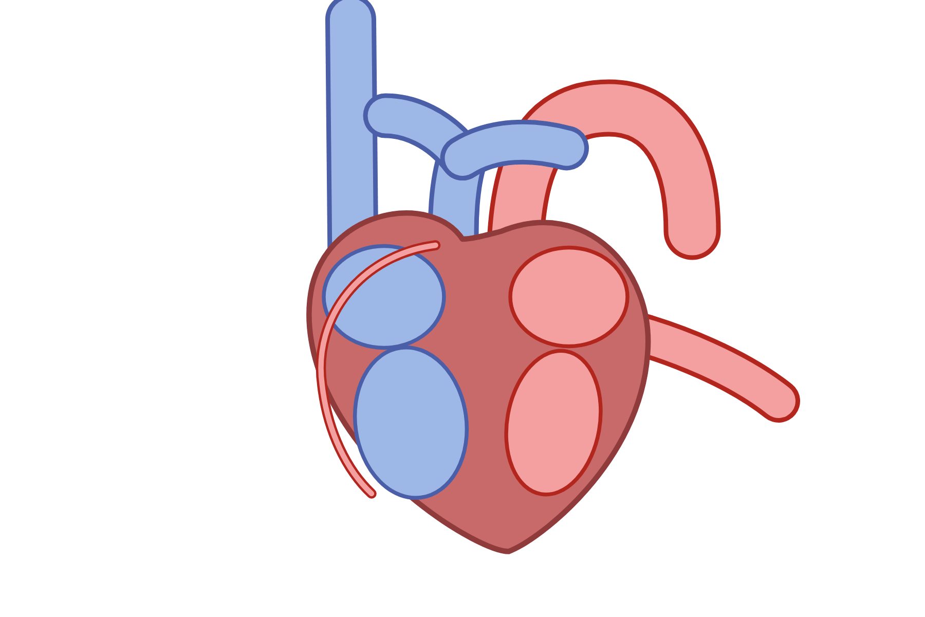 Label every part of the heart.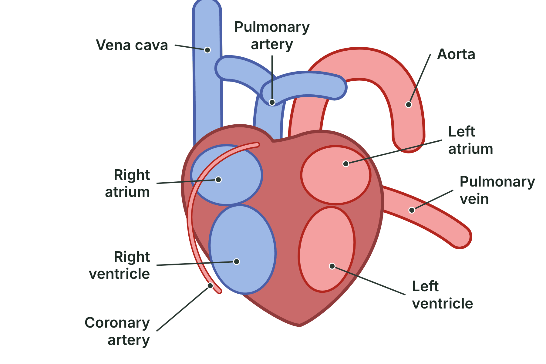
Right atrium, right ventricle, left atrium, left ventricle, vena cava, aorta, pulmonary artery, pulmonary vein and coronary artery.
-
What is an artery?
A vessel that carries blood away from the heart.
-
What is a vein?
A vessel that carries blood back to the heart.
-
What is a capillary?
A tiny vessel with a wall one cell thick, where exchange happens.
-
Why do arteries have thick walls?
The blood is at high pressure, so the walls must be strong and able to stretch and recoil.
-
What are in an artery wall to help it stretch?
Muscle and elastic fibres.
-
Why do veins have valves?
To stop the low-pressure blood flowing backwards.
-
Why is a vein's lumen wide?
It gives less resistance to the flow of low-pressure blood.
-
Why is a capillary wall only one cell thick?
It gives a short diffusion path for exchanging substances.
-
Why do capillaries form huge networks?
They give a large surface area for exchange with the cells.
-
Does an artery always carry oxygenated blood?
No. The pulmonary artery carries deoxygenated blood to the lungs.
-
Does a vein always carry deoxygenated blood?
No. The pulmonary vein carries oxygenated blood from the lungs.
-
What is the formula for rate of blood flow?
Rate = volume of blood ÷ time.
-
600 cm³ of blood flows through a vessel in 2 minutes. What is the rate?
600 ÷ 2 = 300 cm³ per minute.
-
Which vessel type can you feel a pulse in?
An artery.
-
Why does blood move slowly through capillaries?
It gives more time for substances to diffuse.
-
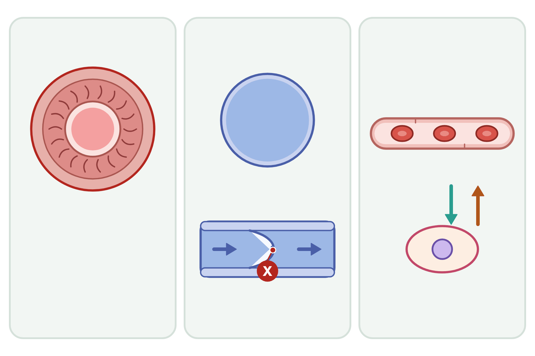 Name the vessel marked X and say how its structure suits its job.
 A valve in a vein. It closes to stop blood flowing backwards, because the blood is at low pressure.
-
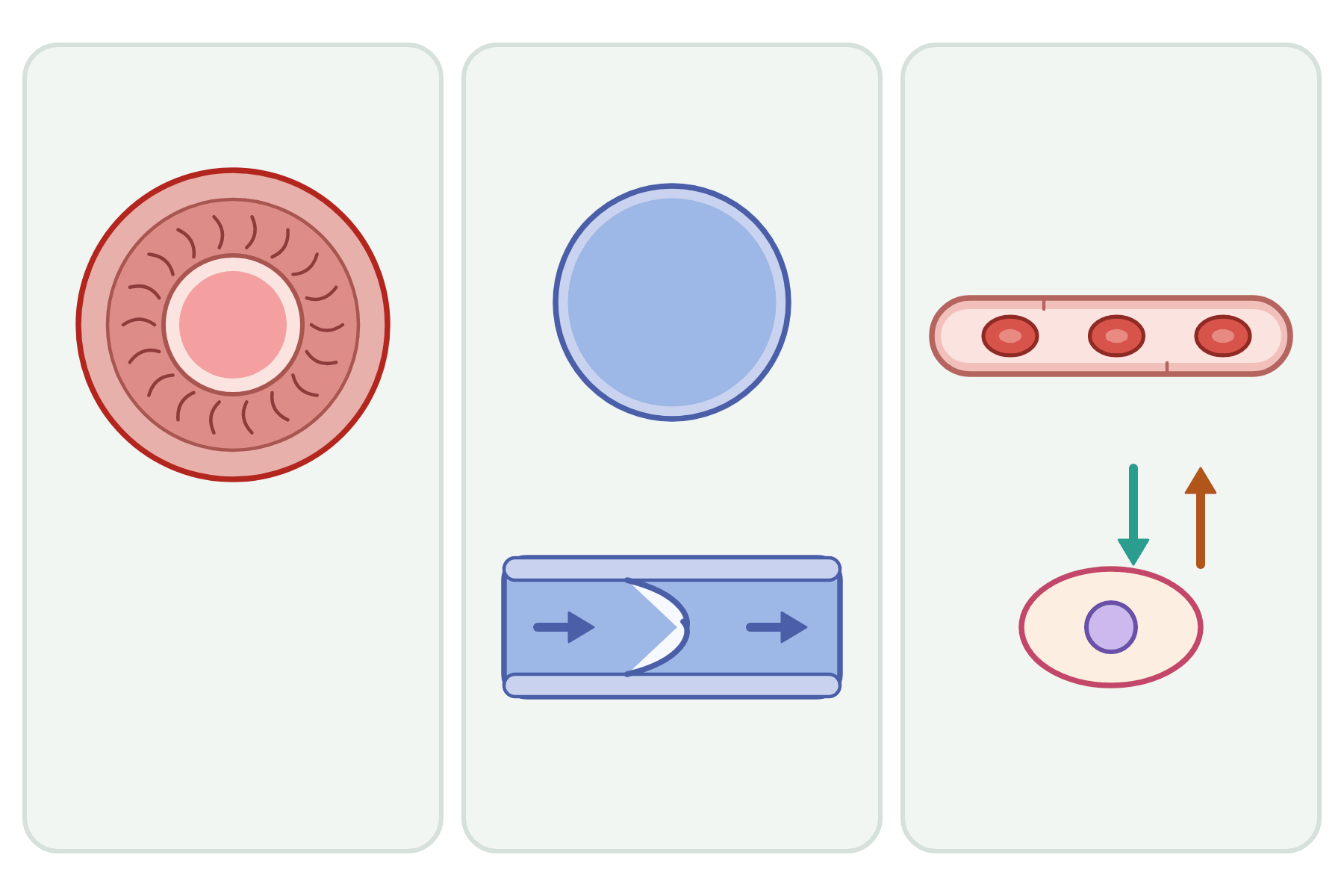 Label the three blood vessels and their features.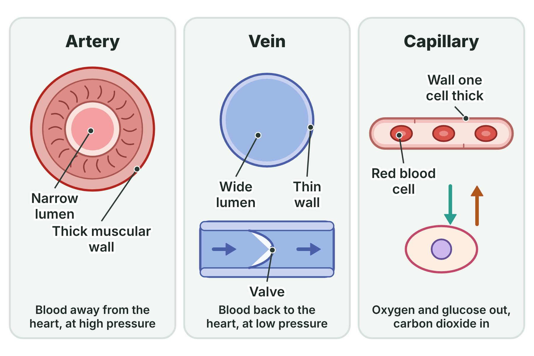
Artery: thick muscular wall, elastic fibres, narrow lumen. Vein: thin wall, wide lumen, valve. Capillary: wall one cell thick.
-
What is the windpipe called?
The trachea.
-
What does the trachea split into?
Two bronchi, one for each lung.
-
What do the bronchioles end in?
Alveoli.
-
What are alveoli?
Tiny air sacs where gas exchange takes place.
-
What surrounds each alveolus?
A network of capillaries.
-
Which gas moves from the alveoli into the blood?
Oxygen.
-
Which gas moves from the blood into the alveoli?
Carbon dioxide.
-
How do the gases move across the alveolus wall?
By diffusion, down their concentration gradients.
-
Why do the lungs have millions of alveoli?
To give a very large surface area for gas exchange.
-
Why are the alveolus walls only one cell thick?
To give a short diffusion path.
-
Why does a rich blood supply help gas exchange?
It carries oxygen away and brings carbon dioxide, so the concentration gradients stay steep.
-
How does breathing help gas exchange?
It ventilates the alveoli, which keeps the concentration gradients steep.
-
What happens to the alveoli in emphysema?
Their walls break down, so the surface area for gas exchange is reduced.
-
How do you work out the volume of air breathed per minute?
Volume per breath × number of breaths per minute.
-
Is air breathed out the same as air breathed in?
No. It has less oxygen and more carbon dioxide.
-
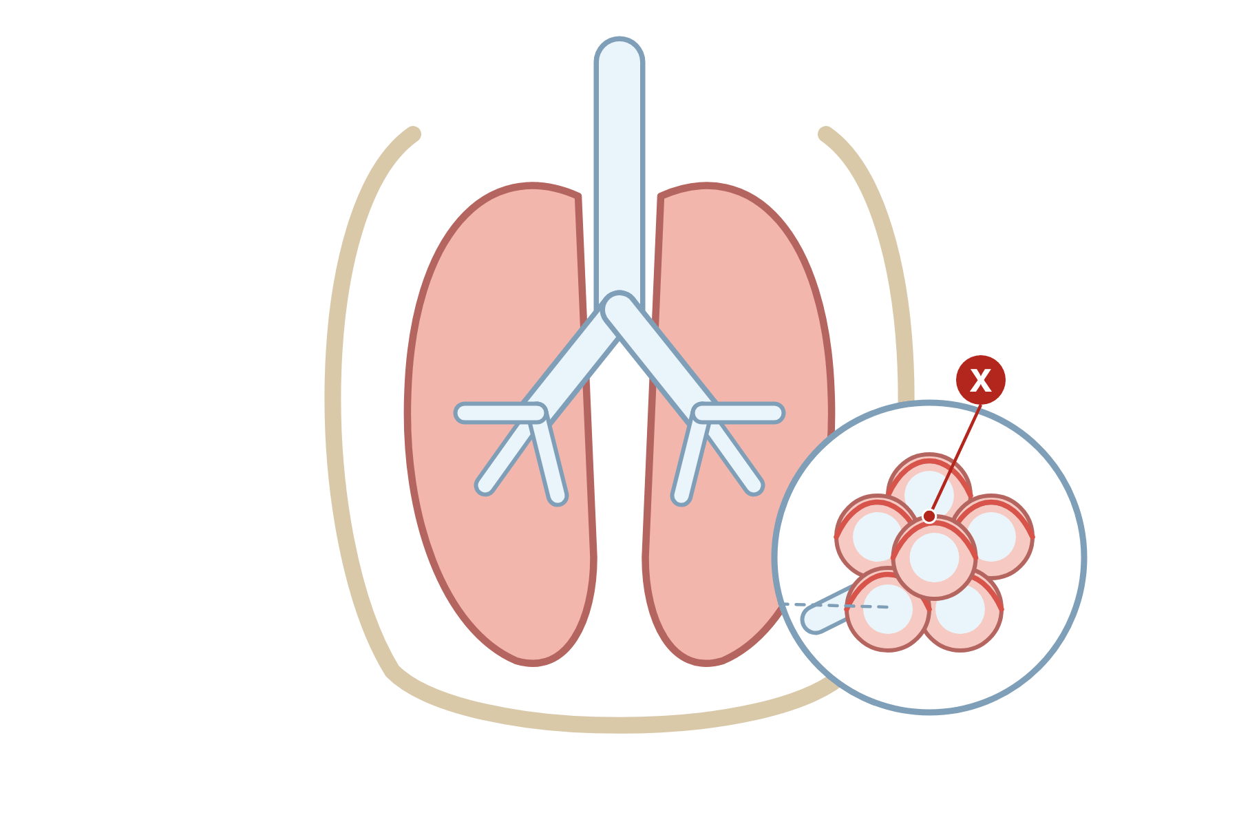 Name the part marked X and say what happens there.
 The alveoli. Oxygen diffuses into the blood and carbon dioxide diffuses out here.
-
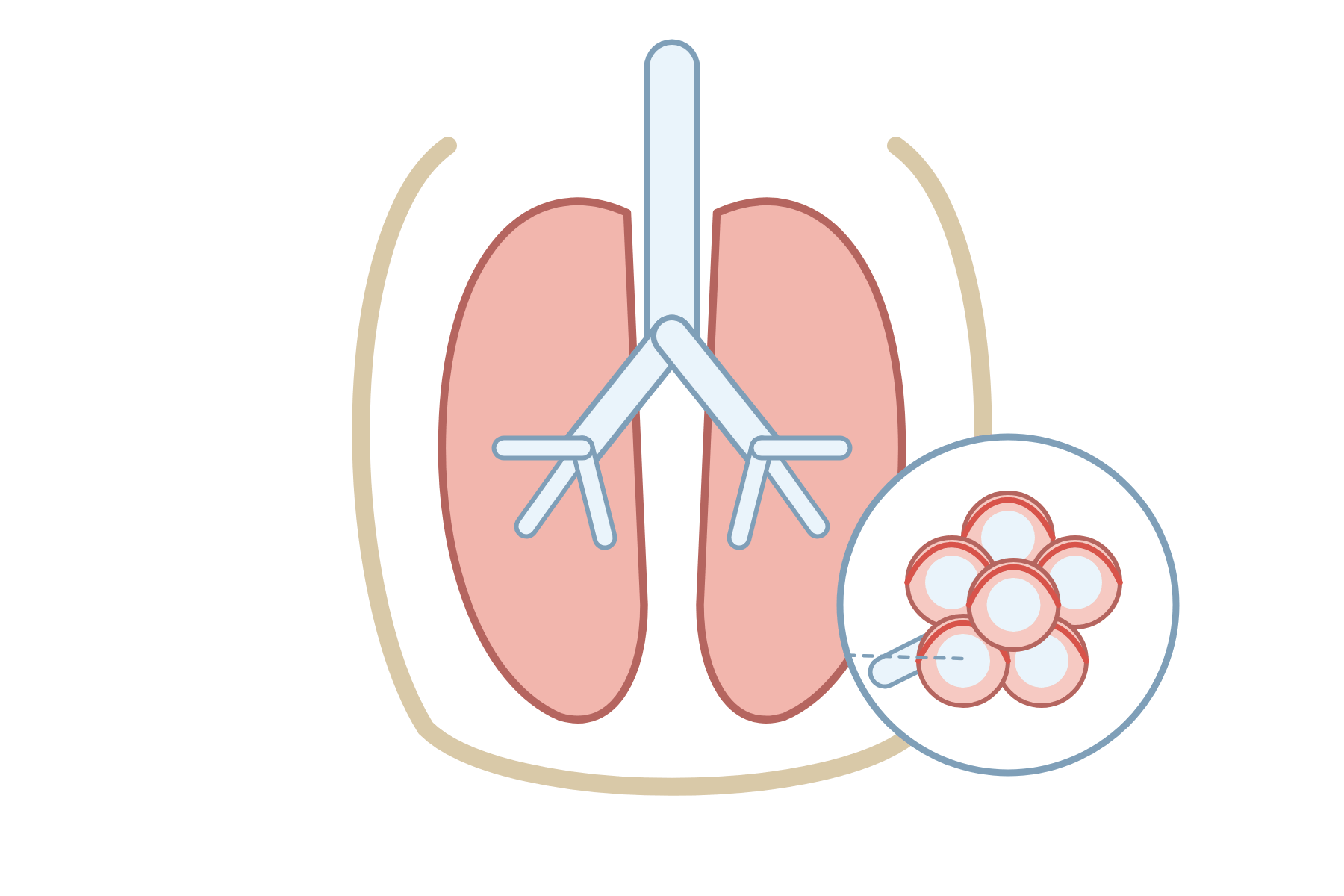 Label the airways and the structures of the lungs.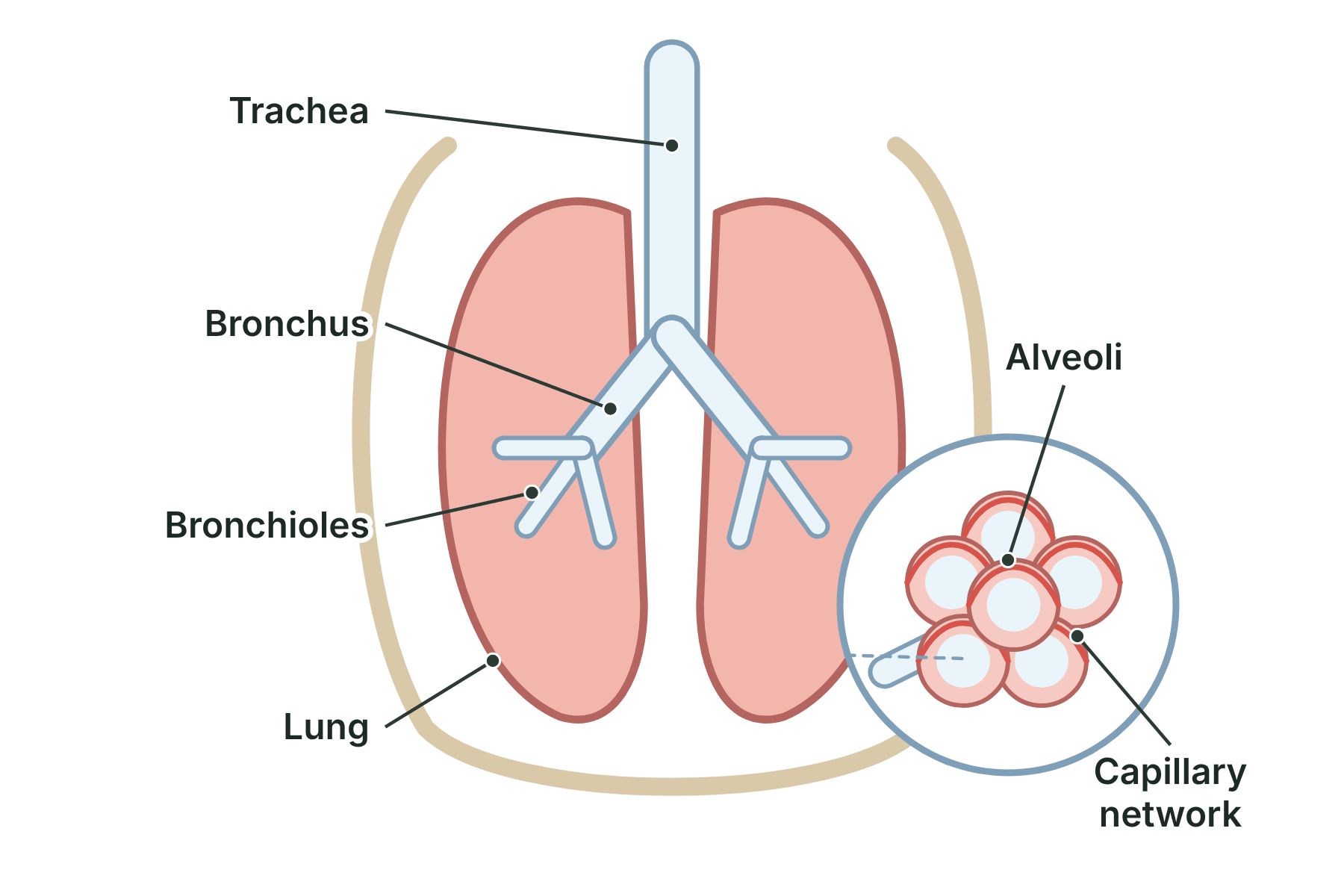
Trachea, bronchus, bronchioles, lung, alveoli and capillary network.
-
What are the four components of blood?
Plasma, red blood cells, white blood cells and platelets.
-
What is plasma?
The pale yellow liquid part of the blood that carries dissolved substances.
-
Name three substances transported in plasma.
Carbon dioxide, soluble food molecules such as glucose, and urea (also hormones and heat).
-
What do red blood cells do?
They carry oxygen from the lungs to the body cells.
-
What is haemoglobin?
The red pigment in red blood cells that binds to oxygen.
-
Why are red blood cells biconcave?
The shape gives a large surface area for oxygen to diffuse in and out quickly.
-
Why do red blood cells have no nucleus?
There is more room for haemoglobin, so they can carry more oxygen.
-
What do white blood cells do?
They help to defend the body against pathogens.
-
Do white blood cells have a nucleus?
Yes.
-
What are platelets?
Small fragments of cells that help the blood to clot.
-
What do platelets do at a wound?
They cause the blood to clot, forming a scab that stops bleeding and keeps microorganisms out.
-
How can you tell a white blood cell from a red blood cell in a photograph?
The white blood cell is larger and has a stained nucleus. The red blood cell has no nucleus.
-
What carries urea in the blood?
The plasma.
-
Give one risk of a blood transfusion.
Infection from the donor blood, a mismatched blood group or an allergic reaction.
-
Which part of the blood carries heat energy?
The plasma.
-
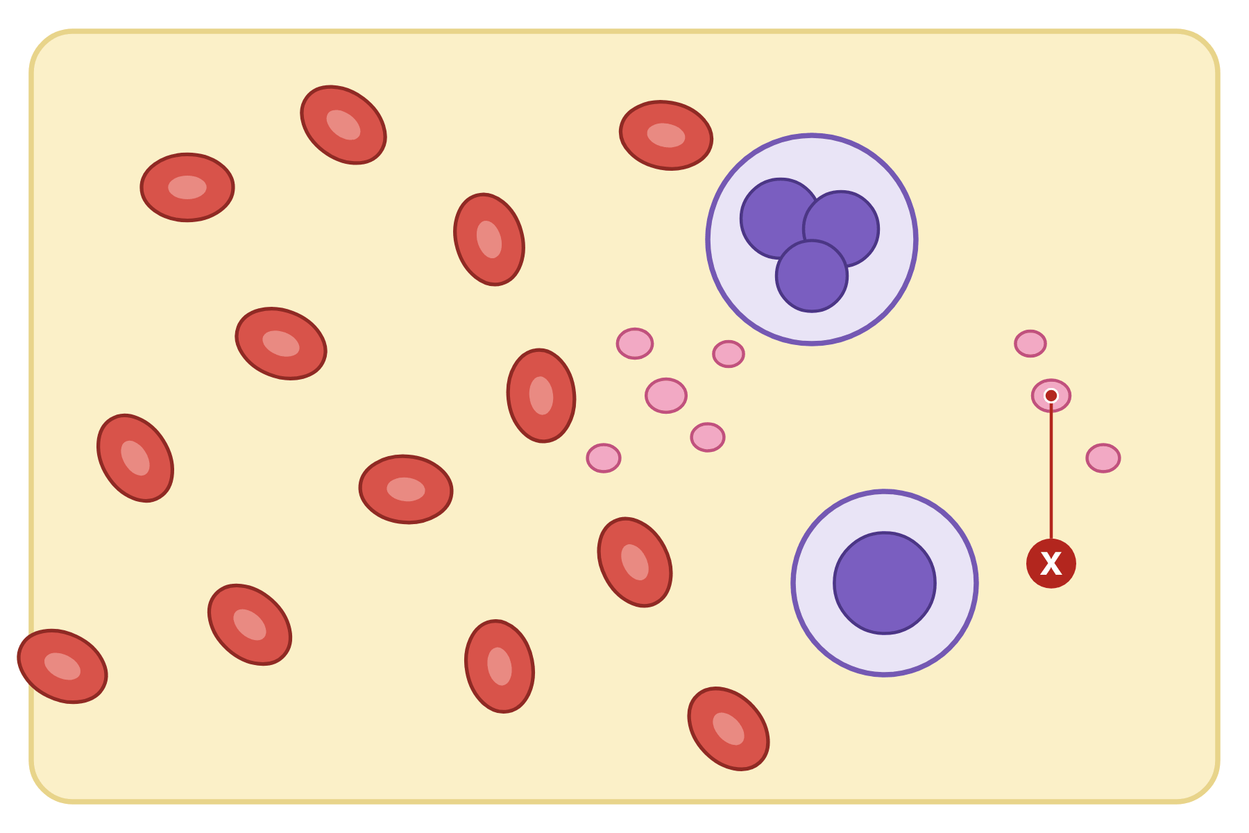 Name the blood component marked X and say what it does.
 A platelet. It is a small fragment of a cell that helps the blood to clot at a wound.
-
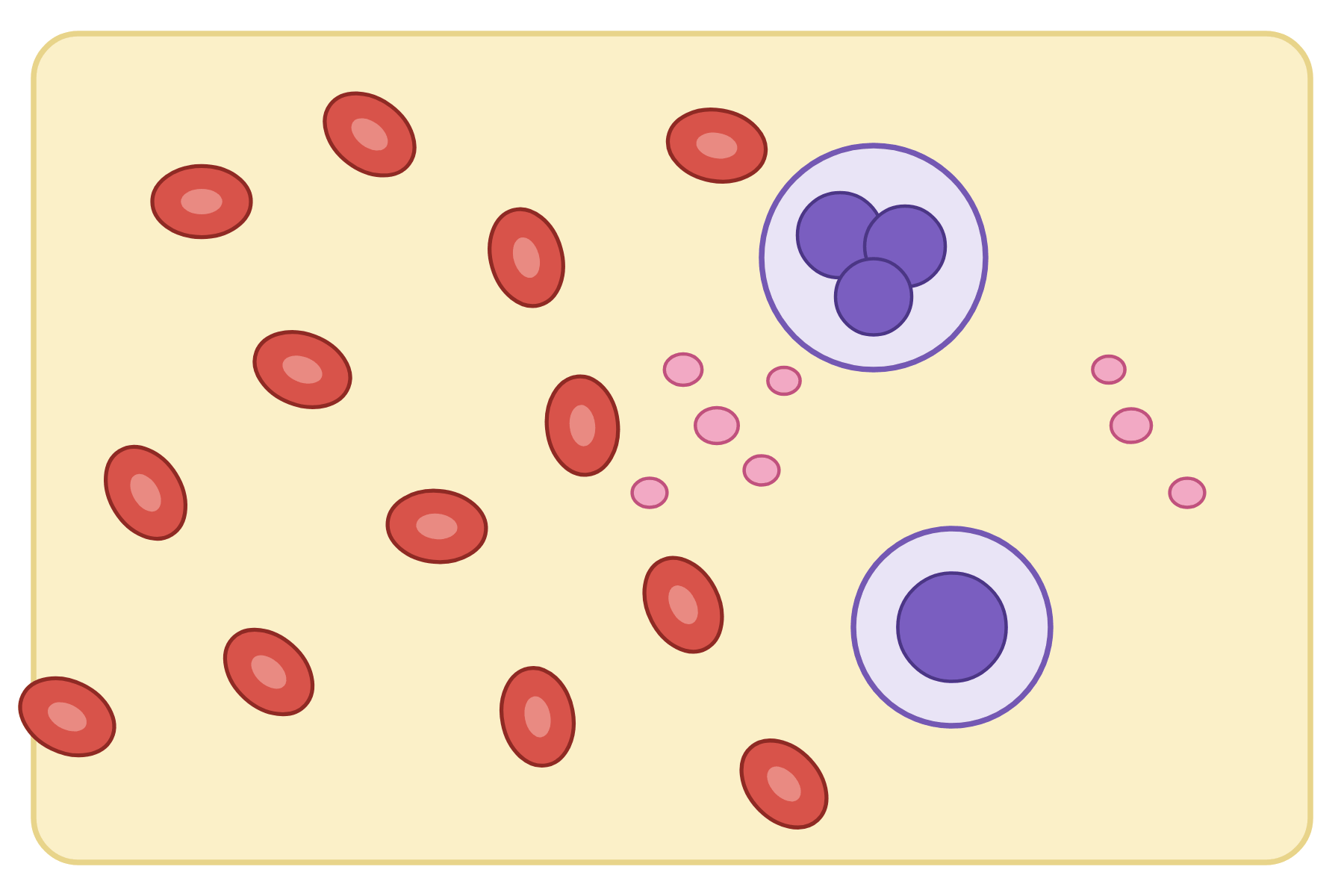 Label every component of the blood.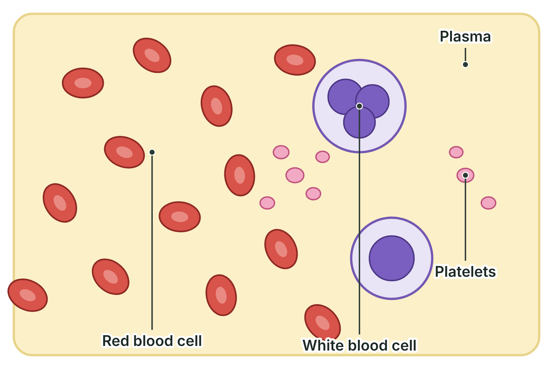
Plasma, red blood cell (biconcave, no nucleus), white blood cell (has a nucleus) and platelets.
-
What is coronary heart disease?
A disease in which layers of fatty material build up inside the coronary arteries and narrow them.
-
Why is coronary heart disease a non-communicable disease?
It is not caused by a pathogen, so it cannot be passed from one person to another.
-
What happens when the coronary arteries narrow?
Less blood reaches the heart muscle, so it gets too little oxygen.
-
What is a stent?
A wire mesh tube put in a narrowed artery to hold it open.
-
What is a statin?
A drug that lowers blood cholesterol, which slows down the build-up of fatty deposits.
-
Give one advantage of a stent.
It keeps the artery open straight away, so blood flows freely again.
-
Give one disadvantage of a stent.
It needs surgery, and the artery can narrow again.
-
Give one disadvantage of statins.
They must be taken for a long time and can have side effects.
-
What can go wrong with a heart valve?
It may not open fully, or it may leak.
-
What can replace a faulty heart valve?
A biological valve or a mechanical valve.
-
Give one advantage of a mechanical valve.
It lasts a long time.
-
Give one disadvantage of a mechanical valve.
The patient needs drugs to stop the blood clotting.
-
When is a heart transplant used?
In heart failure, to replace the diseased heart with a donor heart (or heart and lungs).
-
Give two problems with heart transplants.
There are not enough donors, and the patient must take drugs for life to prevent rejection.
-
Why are artificial hearts used?
To keep a patient alive while they wait for a transplant, or to let the heart rest while it recovers.
-
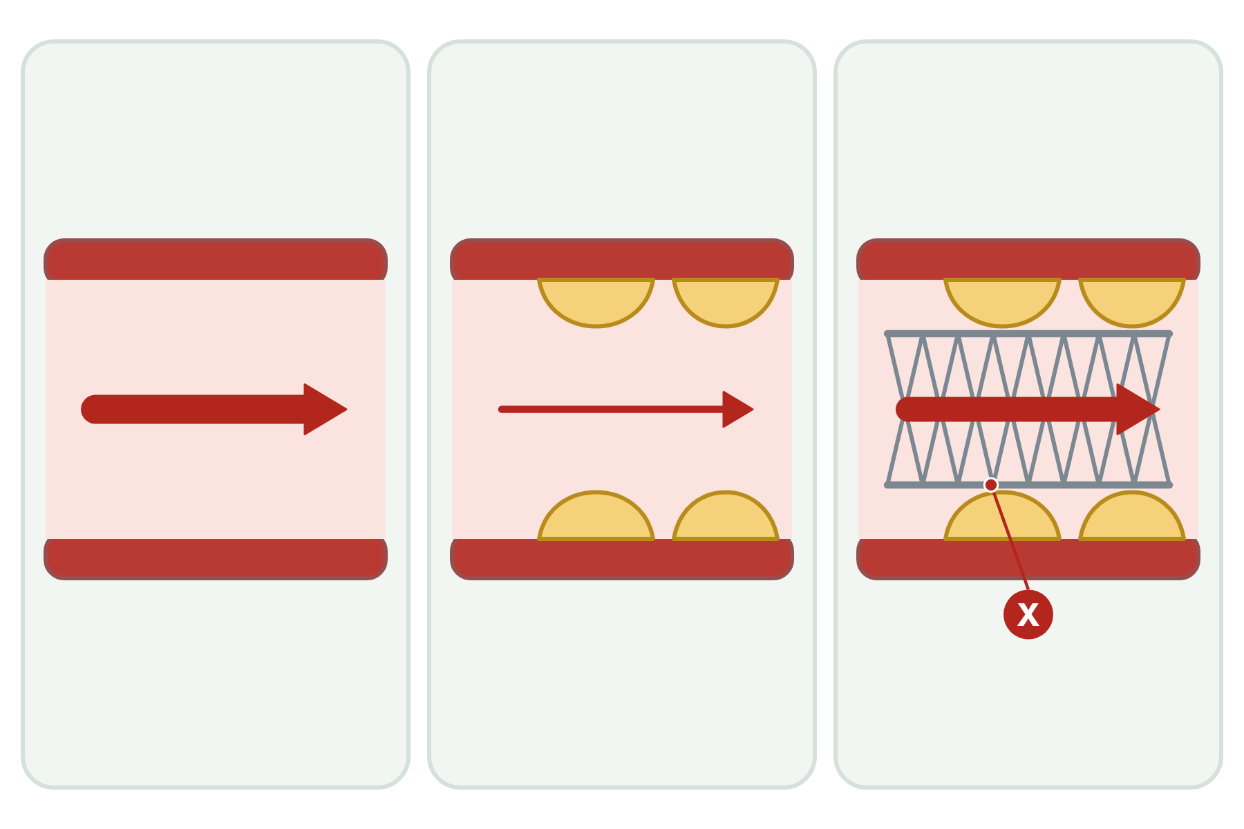 Name the structure marked X in this treated artery and say what it does.
 A stent. It is a mesh tube that holds the narrowed artery open so that blood can flow to the heart muscle.
-
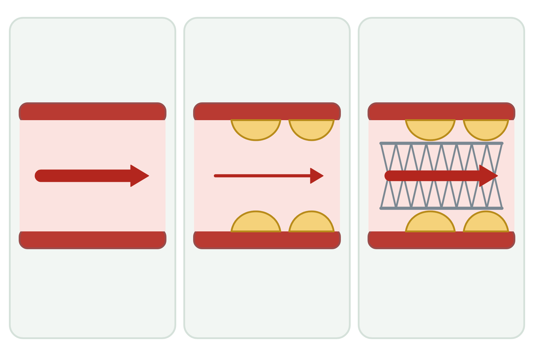 Label the three arteries and the features that matter.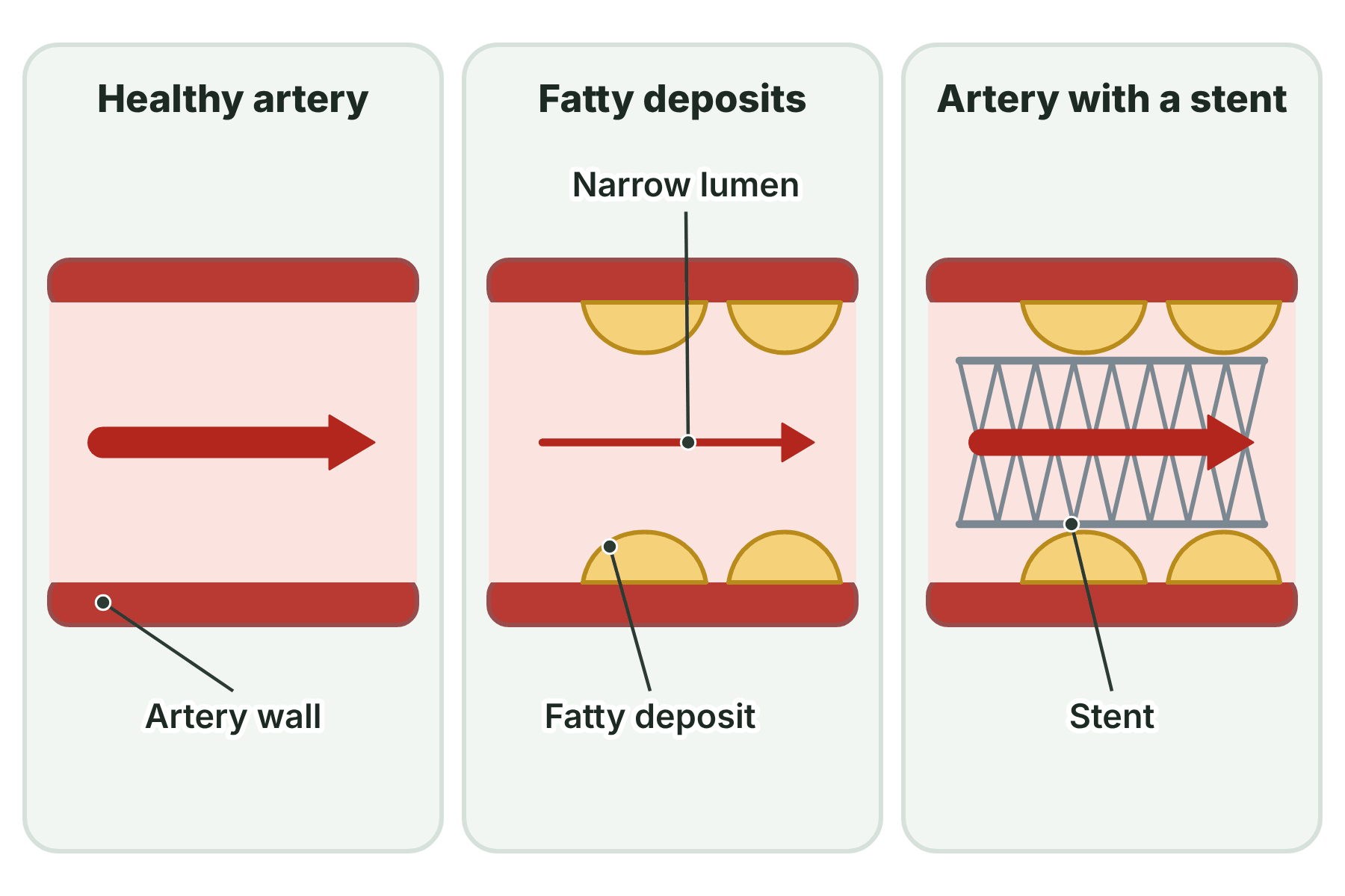
Healthy artery with a wide lumen. Artery wall with fatty deposit and narrow lumen. Artery with a stent holding the lumen open. Blood flow is restored.