Exam questions · Biology · Cell Structure
Microscopy and Magnification
- 8 exam questions
- 21 marks
- 8 quick checks
-
1 State [1 mark]
State the formula used to calculate magnification.
Show answerHide answer
Model answer
Magnification = size of image ÷ size of real object.
Mark scheme
- Magnification = image size ÷ real size — 1 mark
-
2 Give [2 marks]
Give two advantages of using an electron microscope instead of a light microscope.
Show answerHide answer
Model answer
It has a higher magnification, and it has a higher resolution so more detail can be seen.
Mark scheme
- Higher magnification — 1 mark
- Higher resolution / shows more detail — 1 mark
-
3 Calculate [3 marks]
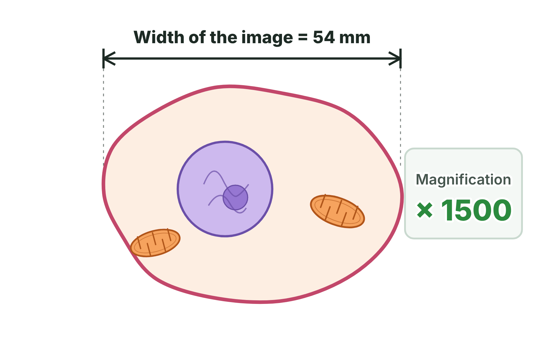
Figure 1 shows a drawing of a cell. The magnification is ×1500. Calculate the real width of the cell. Give your answer in micrometres.
Show answerHide answer
Model answer
real size = 54 ÷ 1500 = 0.036 mm. 0.036 mm × 1000 = 36 µm.
Mark scheme
- Uses real size = image size ÷ magnification — 1 mark
- 54 ÷ 1500 = 0.036 mm — 1 mark
- Converts to 36 µm — 1 mark
-
4 Calculate [2 marks]
A student looks at a slide using a ×10 eyepiece lens and a ×40 objective lens. Calculate the total magnification, and name the wheel the student should use to make the image sharp.
Show answerHide answer
Model answer
Total magnification = 10 × 40 = ×400. The student should use the fine focus wheel.
Mark scheme
- 10 × 40 = ×400 — 1 mark
- Fine focus wheel — 1 mark
-
5 Describe [4 marks]
Describe how to use a light microscope to look at a slide of onion cells.
Show answerHide answer
Model answer
Place the slide on the stage and secure it with the clips. Select the lowest power objective lens. Use the coarse focus wheel to bring the image roughly into focus, looking from the side first so the lens does not hit the slide. Then use the fine focus wheel to make the image sharp. Change to a higher power lens and refocus with the fine wheel if more detail is needed.
Mark scheme
- Slide on the stage, held by clips — 1 mark
- Start with the lowest power objective lens — 1 mark
- Coarse focus to find the image — 1 mark
- Fine focus to sharpen it — 1 mark
-
6 Explain [4 marks]
Explain how electron microscopy has increased our understanding of sub-cellular structures.
Show answerHide answer
Model answer
An electron microscope has a much higher magnification and resolving power than a light microscope. This means it can show structures that are too small to be seen with a light microscope, such as ribosomes, plasmids and the internal structure of mitochondria and chloroplasts. Scientists have used this detail to work out how the structures carry out their functions.
Mark scheme
- Electron microscopes have higher magnification — 1 mark
- Electron microscopes have higher resolution / resolving power — 1 mark
- Shows structures not visible with a light microscope, for example ribosomes or the inside of mitochondria — 1 mark
- Understanding of how structures work (their functions) has improved — 1 mark
-
7 Calculate [2 marks]
An electron micrograph shows a chloroplast that is 25 mm long. The real chloroplast is 5 µm long. Calculate the magnification. Give your answer in standard form.
Show answerHide answer
Model answer
25 mm = 25 000 µm. Magnification = 25 000 ÷ 5 = 5000, which is \(5 \times 10^{3}\).
Mark scheme
- Converts 25 mm to 25 000 µm and divides by 5 — 1 mark
- \(5 \times 10^{3}\) — 1 mark
-
8 Calculate [3 marks]
A ribosome is 25 nm across. In an electron micrograph the image of the ribosome is 5 mm across. Calculate the magnification. Give your answer in standard form.
Show answerHide answer
Model answer
5 mm = 5 000 000 nm. Magnification = 5 000 000 ÷ 25 = 200 000, which is \(2 \times 10^{5}\).
Mark scheme
- Converts 5 mm to 5 000 000 nm — 1 mark
- 5 000 000 ÷ 25 = 200 000 — 1 mark
- \(2 \times 10^{5}\) — 1 mark
Quick check
-
1
What does magnification tell you?
Show answerHide answer
B: How many times bigger the image is than the real object
Magnification is how many times bigger the image is than the real object.
-
2
Which microscope has the higher resolution?
Show answerHide answer
C: An electron microscope
An electron microscope has much higher resolving power than a light microscope, so it shows more detail.
-
3
A cell is 2 mm wide in a drawing. The real cell is 0.02 mm wide. What is the magnification?
Show answerHide answer
D: ×100
Magnification = image size ÷ real size = 2 ÷ 0.02 = 100, so ×100.
-
4
Which of these can a light microscope show?
Show answerHide answer
A: A chloroplast
A light microscope shows nuclei, cell walls and chloroplasts. Ribosomes and plasmids are too small and need an electron microscope.
-
5
Which lens should you use first when focusing a specimen?
Show answerHide answer
B: The lowest power objective lens
Start with the lowest power lens so that you can find the specimen in a wide field of view.
-
6
How many micrometres are there in 1 mm?
Show answerHide answer
C: 1000
Milli and micro differ by a factor of 1000, so 1 mm is 1000 µm.
-
7
A drawing shows a cell as 45 mm wide. The cell is really 15 µm wide. What is the magnification?
Show answerHide answer
D: ×3000
Convert 45 mm to 45 000 µm, then divide by 15 µm to get 3000.
-
8
An image is 6 mm wide and the magnification is ×200. What is the real width?
Show answerHide answer
B: 30 µm
Real size = image size ÷ magnification = 6 ÷ 200 = 0.03 mm, which is 30 µm.