Flashcards · Biology
Homeostasis
-
What is homeostasis?
The regulation of the internal conditions of the body to keep them constant.
-
Name three conditions that the body controls.
Body temperature, blood glucose concentration and water content.
-
At what temperature is the human body kept?
About 37 °C.
-
Why must body temperature stay constant?
Enzymes work best at an optimum temperature.
-
What happens to enzymes if the body gets too hot?
They denature and stop working.
-
What does a receptor do?
Detects a change in a condition.
-
What does a coordination centre do?
Receives the information and decides on the response.
-
What does an effector do?
Carries out the response.
-
Give two examples of coordination centres.
The brain and the pancreas.
-
What is negative feedback?
A response that reverses a change and brings the condition back to normal.
-
What happens if the condition is too high in negative feedback?
The response makes it fall.
-
What happens if the condition is too low in negative feedback?
The response makes it rise.
-
Why is the water content of the blood controlled?
Osmosis would make cells gain or lose water.
-
Why is blood glucose controlled?
Cells need a supply of glucose for respiration.
-
Is homeostasis automatic?
Yes, you do not have to think about it.
-
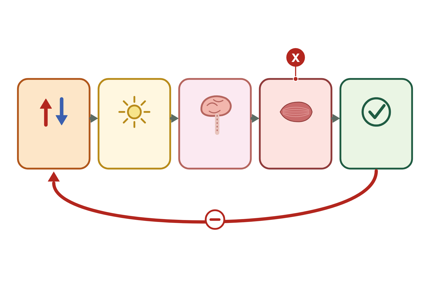 Name the part of the control system marked X.
 The effector. It carries out the response, such as a muscle contracting or a gland releasing a chemical.
-
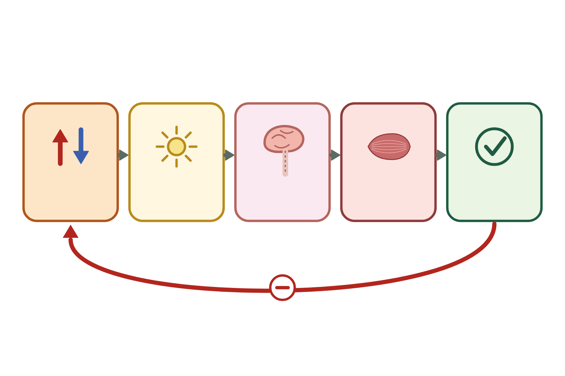 Name each stage of a negative feedback loop.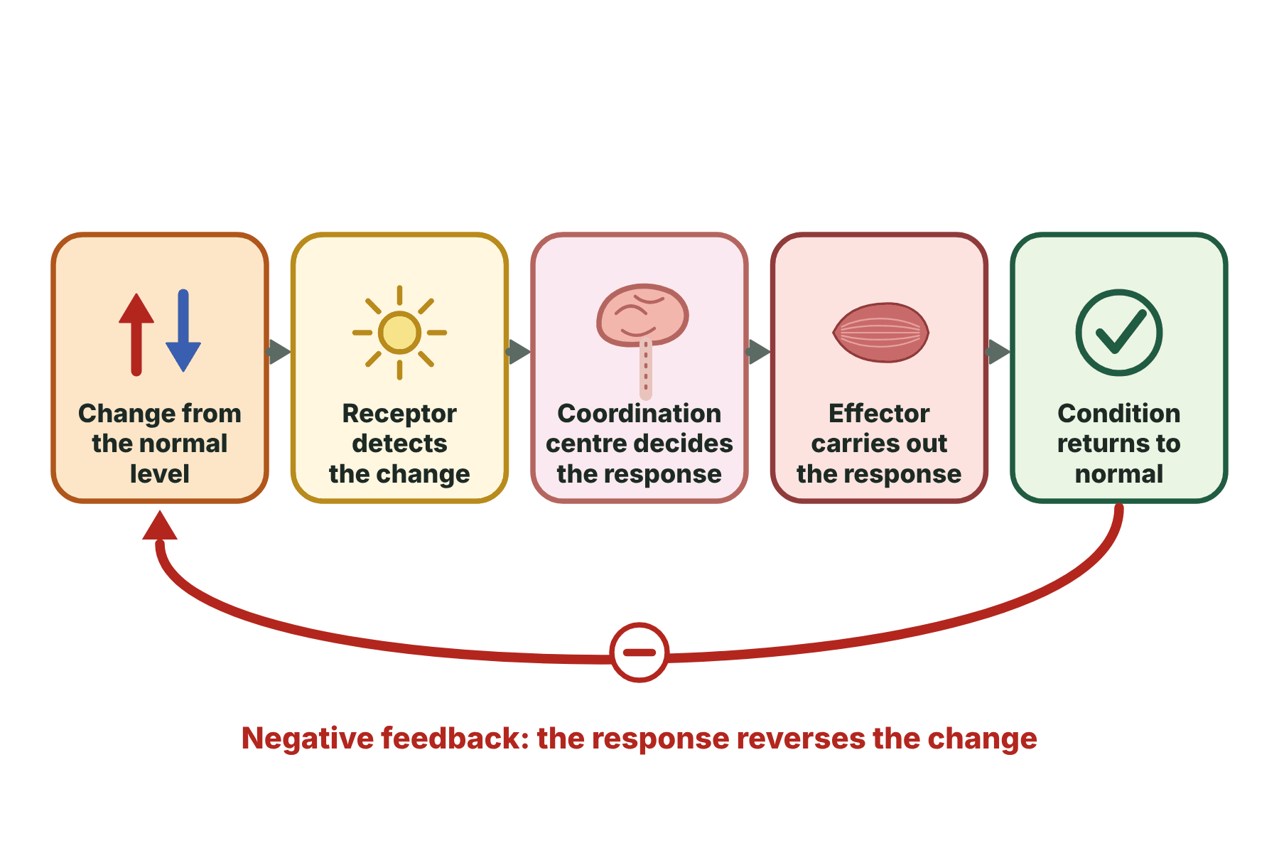
Change from normal, receptor, coordination centre, effector, return to normal. The response reverses the change, which is negative feedback.
-
At what temperature is the body kept?
About 37 °C.
-
Where are temperature receptors found?
In the skin and in the hypothalamus.
-
What is the coordination centre for temperature control?
The hypothalamus in the brain.
-
Name the effectors for temperature control.
Sweat glands, muscles in the walls of blood vessels, hair erector muscles and skeletal muscles.
-
How does sweating cool the body?
Sweat evaporates and takes heat from the skin.
-
What is vasodilation?
The widening of blood vessels near the surface of the skin.
-
How does vasodilation help when too hot?
More blood near the surface, so more heat is lost.
-
What is vasoconstriction?
The narrowing of blood vessels near the surface of the skin.
-
How does vasoconstriction help when too cold?
Less blood near the surface, so less heat is lost.
-
How does shivering warm the body?
Muscle respiration releases heat.
-
What happens to hairs when it is cold?
They stand up, trapping an insulating layer of air.
-
What does the fat layer do?
It insulates the body.
-
Why does the skin look red when hot?
Blood vessels widen and more blood flows near the surface.
-
Why does the skin look pale when cold?
Blood vessels narrow, so less blood flows near the surface.
-
Why is temperature control an example of negative feedback?
A change causes a response that reverses the change.
-
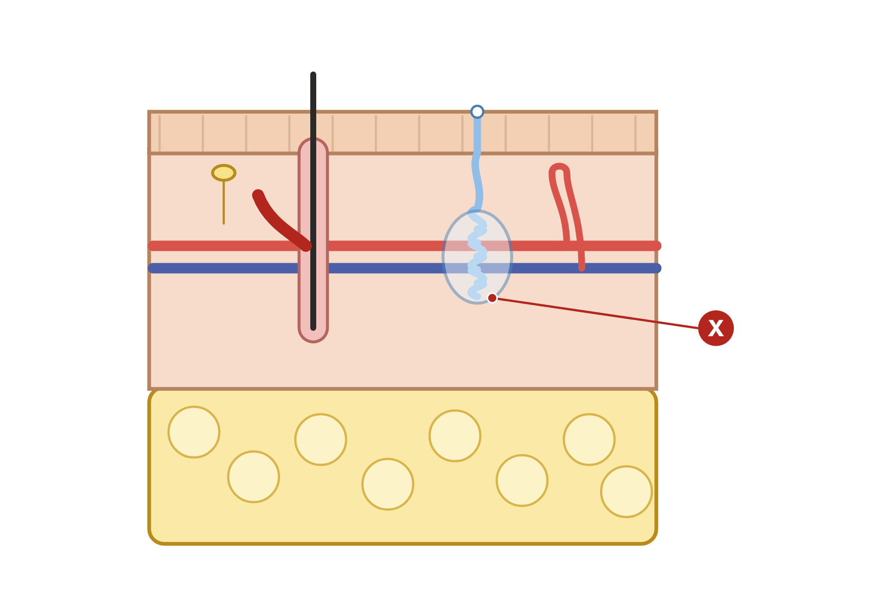 Name the part of the skin marked X and say what it does when it is hot.
 The sweat gland. It releases more sweat, which evaporates from the skin and cools the body.
-
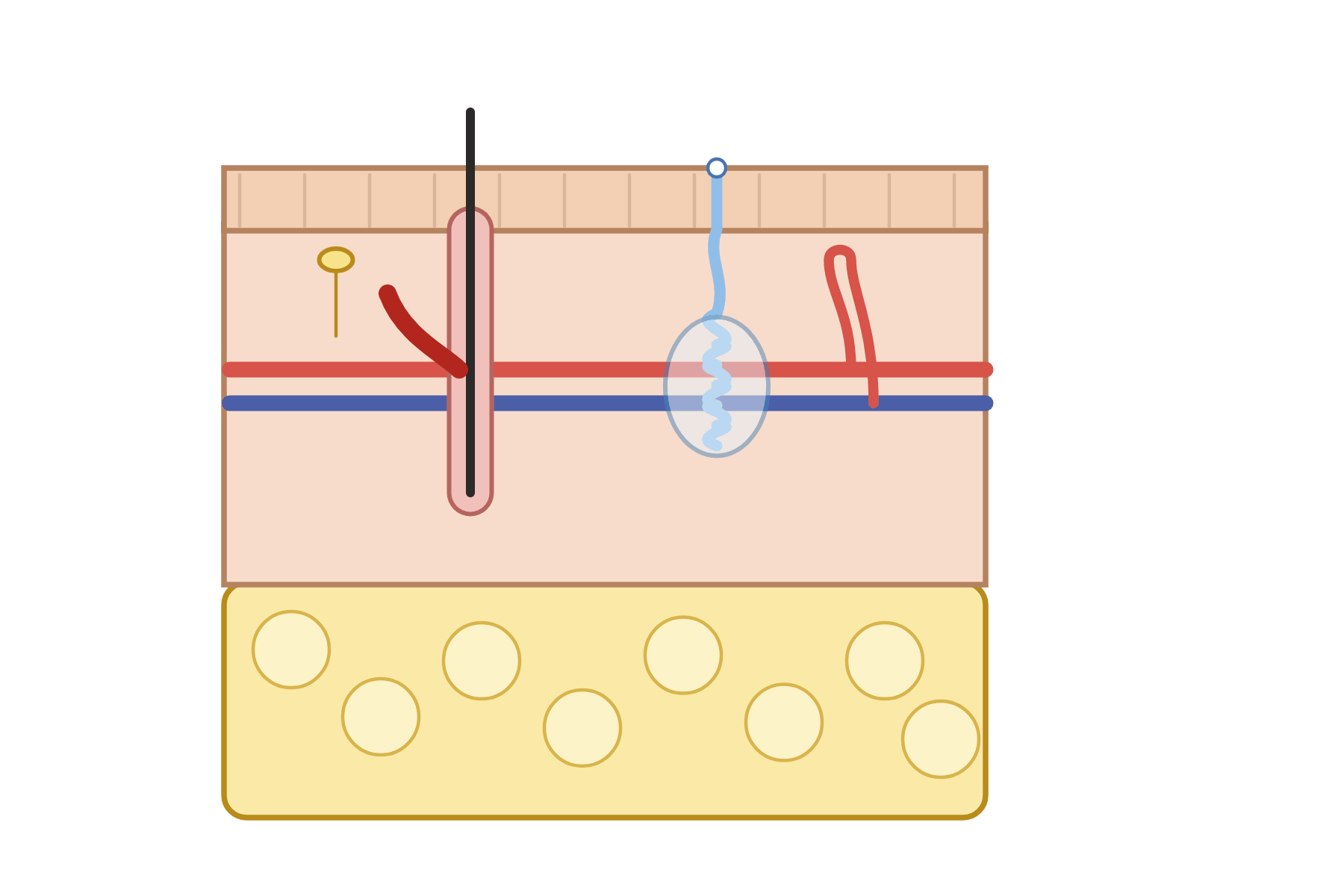 Label the skin.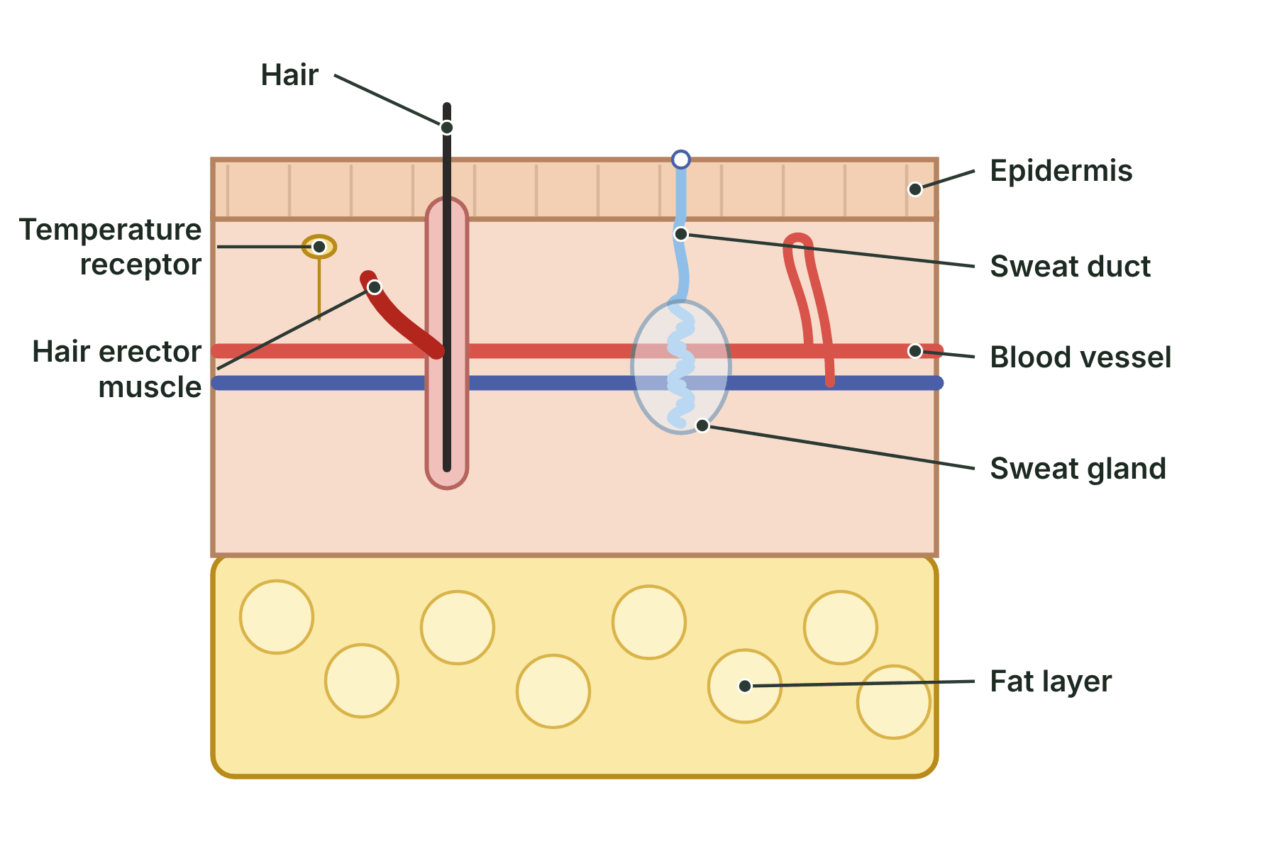
Epidermis, hair, hair erector muscle, sweat gland, sweat duct, blood vessel, temperature receptor and fat layer.
-
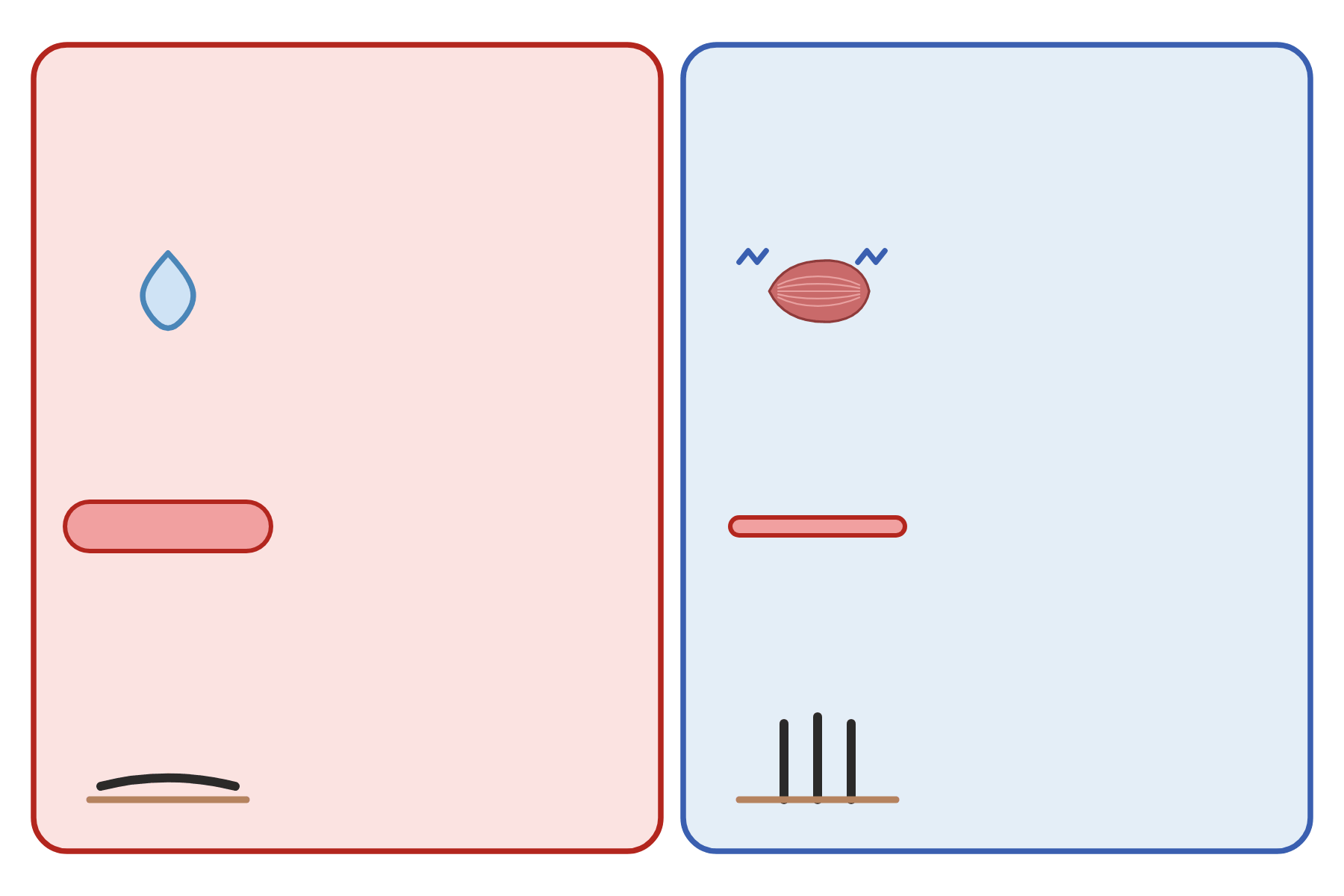 Describe the body's responses to being too hot and too cold.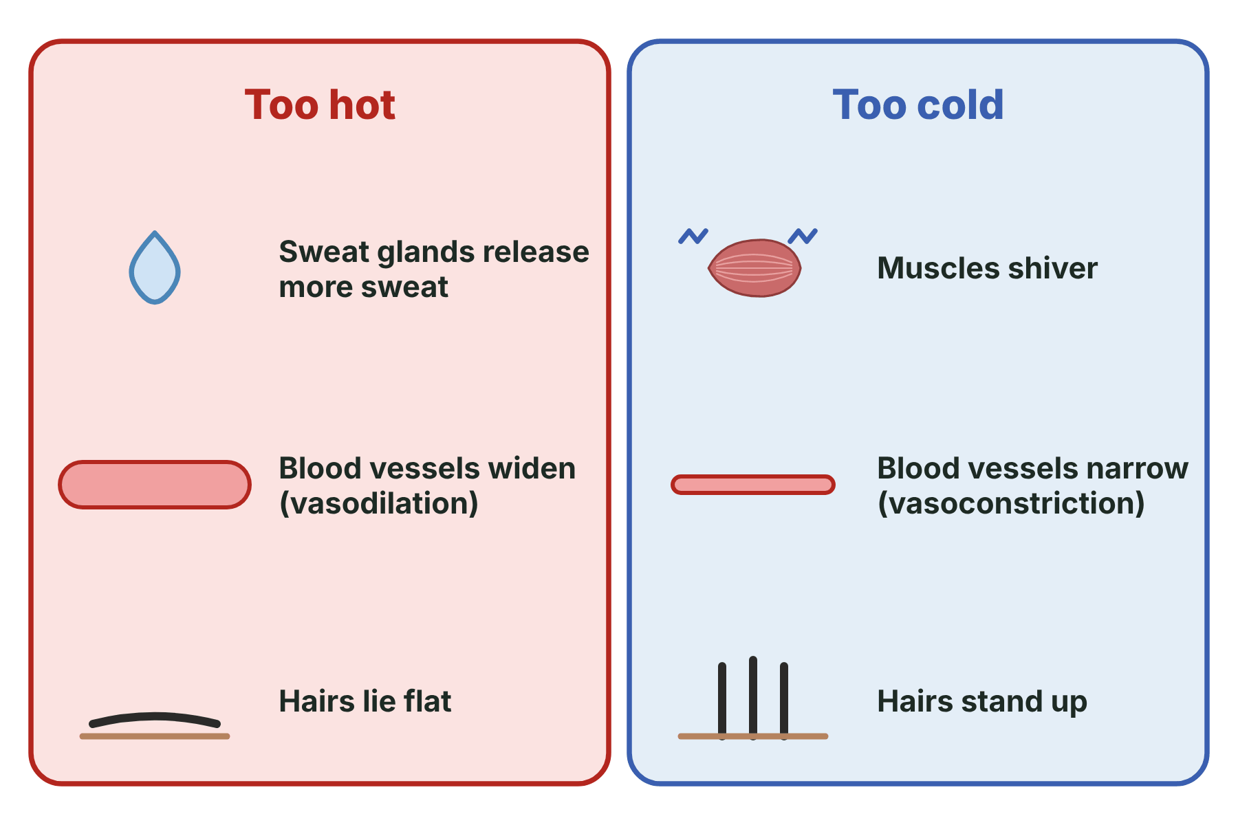
Too hot: more sweat, blood vessels widen, hairs lie flat. Too cold: shivering, blood vessels narrow, hairs stand up.
-
What does insulin do?
Lowers the blood glucose concentration.
-
Where is insulin made?
In the pancreas.
-
What does insulin make the liver do?
Take up glucose and convert it to glycogen.
-
What is glycogen?
The carbohydrate in which the liver stores glucose.
-
What does glucagon do? (Higher tier)
Raises the blood glucose concentration.
-
What does glucagon make the liver do? (Higher tier)
Convert glycogen to glucose and release it.
-
What is diabetes?
A disease in which the blood glucose concentration is not controlled properly.
-
What causes type 1 diabetes?
The pancreas does not make enough insulin.
-
What causes type 2 diabetes?
The body cells stop responding to insulin.
-
How is type 1 diabetes treated?
With insulin injections and a controlled diet.
-
How is type 2 diabetes treated?
With diet, exercise, weight loss and sometimes medicine.
-
Which type of diabetes is linked to obesity?
Type 2.
-
Which type of diabetes usually starts in childhood?
Type 1.
-
Why is blood glucose controlled by negative feedback?
A rise causes insulin release, which makes it fall.
-
Why do people with type 1 diabetes inject insulin?
Their pancreas does not make enough.
-
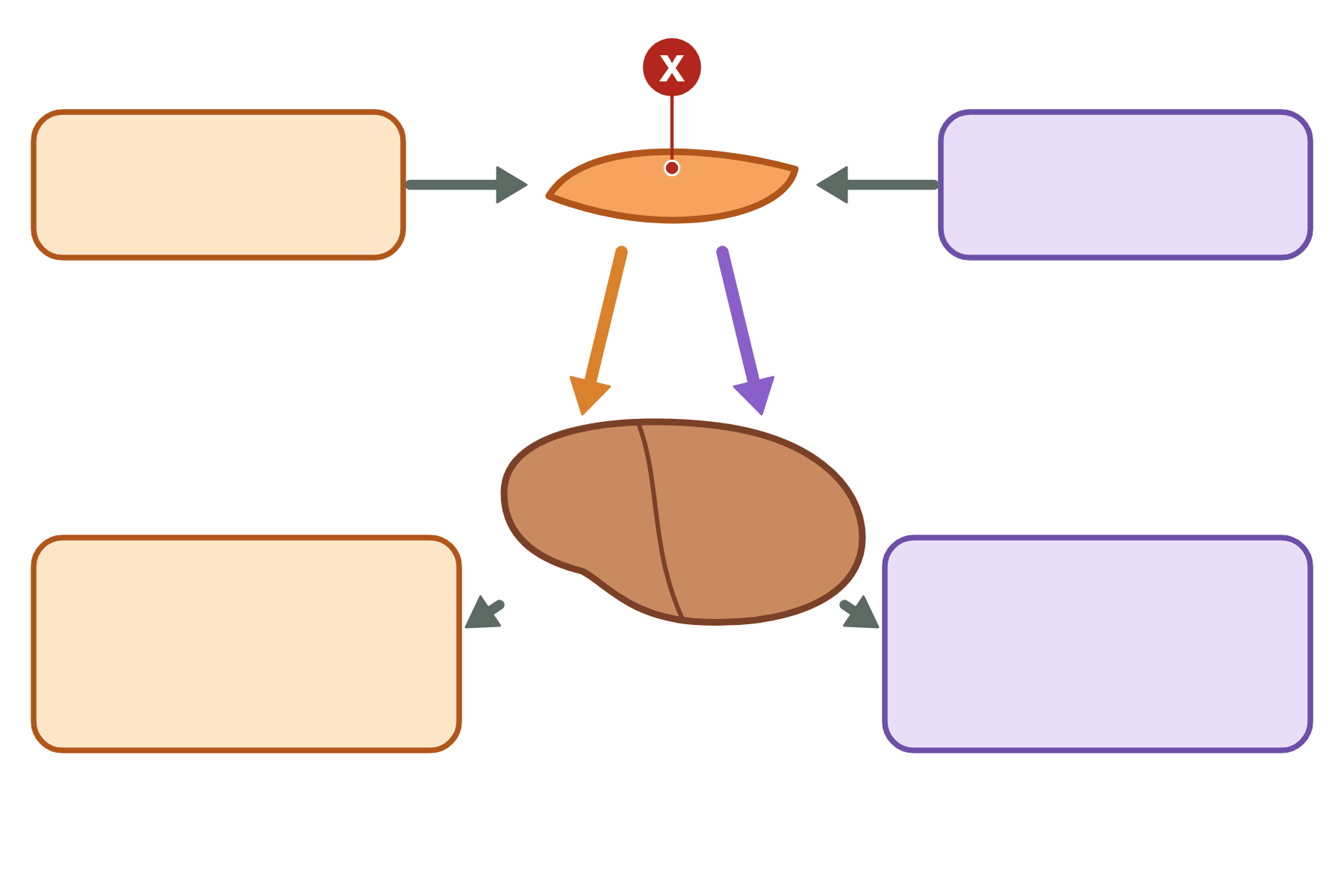 Name the organ marked X and say what it releases.
 The pancreas. It releases insulin, and glucagon (Higher tier).
-
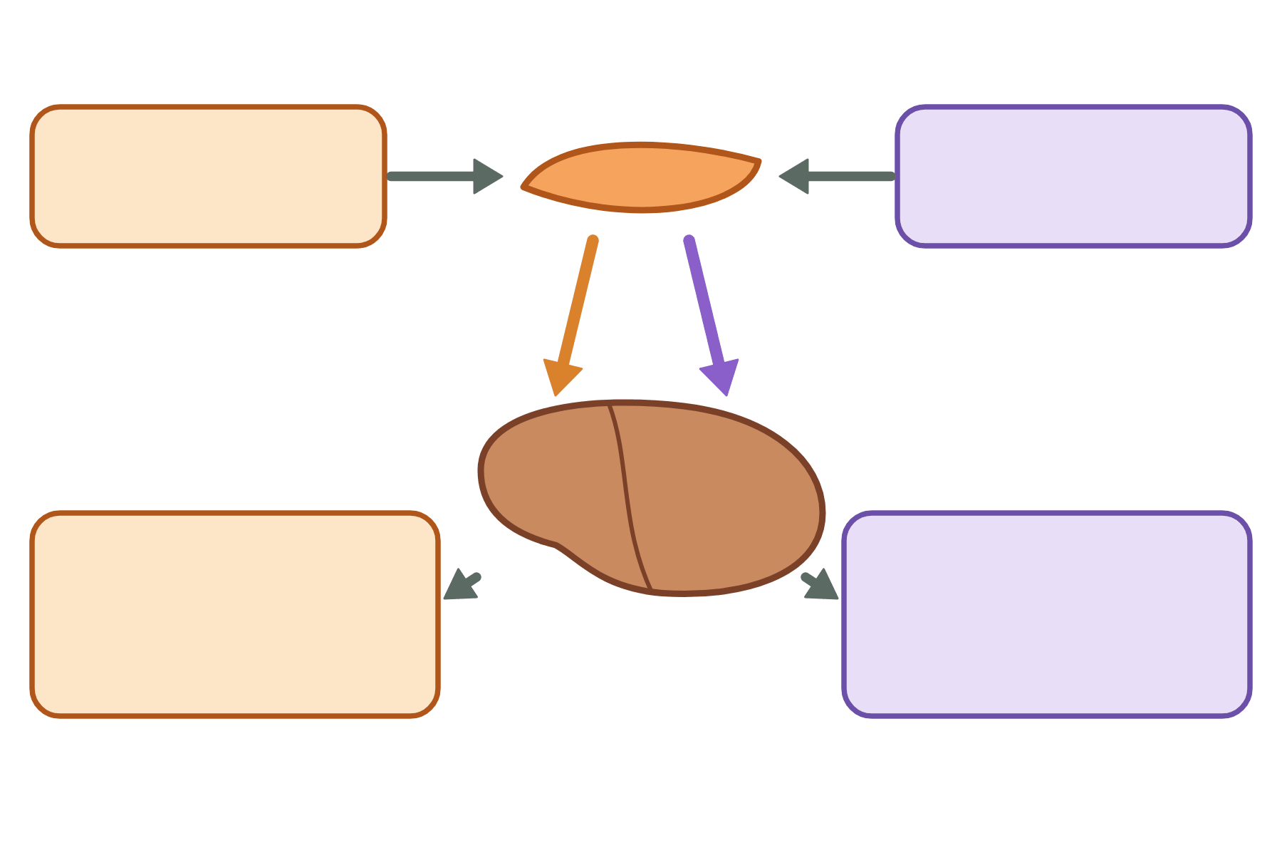 Explain what happens when the blood glucose is too high and too low.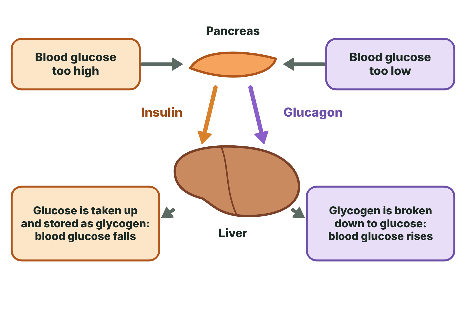
Too high: the pancreas releases insulin, the liver stores glucose as glycogen, and the glucose falls. Too low (Higher tier): the pancreas releases glucagon, the liver releases glucose, and the glucose rises.
-
What happens to an animal cell in a dilute solution?
Water enters by osmosis, so it swells and may burst.
-
What happens to an animal cell in a concentrated solution?
Water leaves by osmosis, so it shrinks.
-
What do the kidneys remove from the blood?
Urea, excess water and excess mineral ions.
-
Where is urea made?
In the liver, from excess amino acids.
-
What carries urine from the kidney to the bladder?
The ureter.
-
What is a nephron?
The tiny filtering unit of the kidney.
-
What is the glomerulus?
A knot of capillaries inside the Bowman's capsule.
-
What passes into the Bowman's capsule during filtration?
Water, glucose, urea and mineral ions.
-
What is not filtered out of the blood?
Blood cells and proteins.
-
What is selective reabsorption?
All the glucose, some water and some ions are taken back into the blood.
-
How is glucose reabsorbed?
By active transport.
-
What is ADH? (Higher tier)
A hormone that controls how much water the kidneys reabsorb.
-
Where is ADH released from? (Higher tier)
The pituitary gland.
-
What does ADH do to the collecting duct? (Higher tier)
Makes it more permeable to water.
-
Why is the urine concentrated after exercise? (Higher tier)
Water is lost in sweat, so more ADH is released and more water is reabsorbed.
-
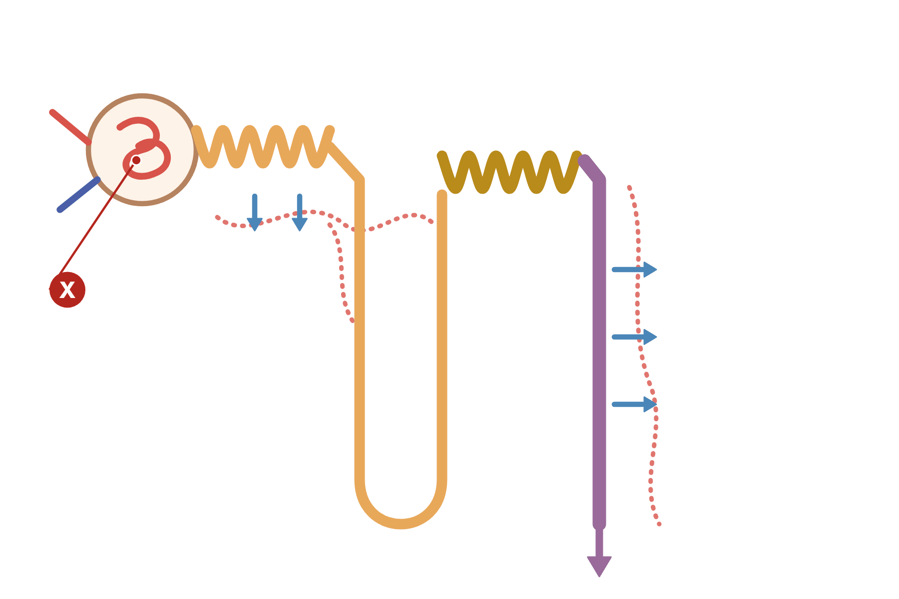 Name the part of the nephron marked X and say what happens there.
 The glomerulus. Blood is filtered here under high pressure, and small molecules pass into the Bowman's capsule.
-
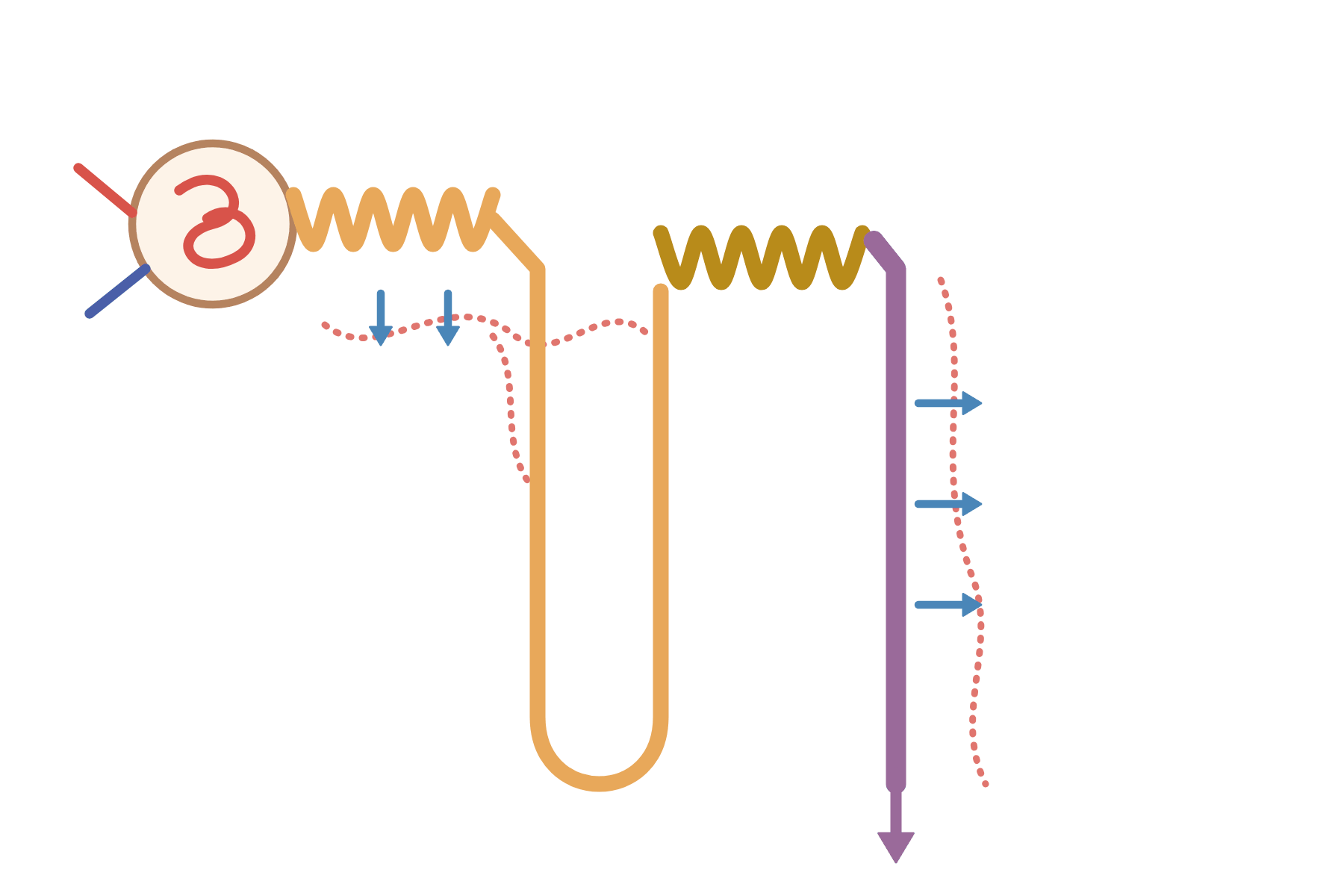 Label the parts of the nephron.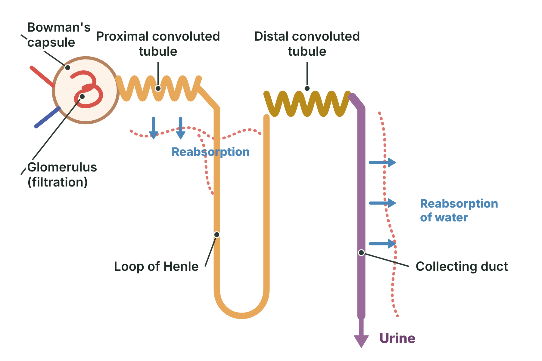
Bowman's capsule, glomerulus, proximal convoluted tubule, loop of Henle, distal convoluted tubule and collecting duct.
-
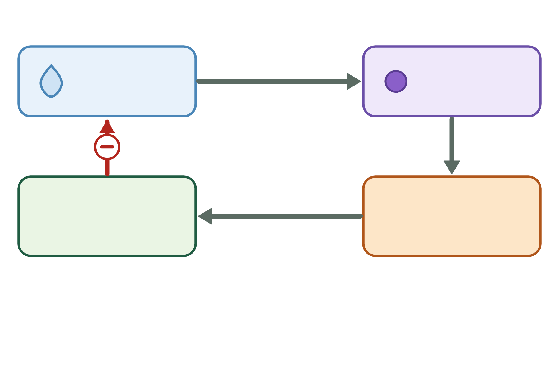 Describe how ADH keeps the water content of the blood constant.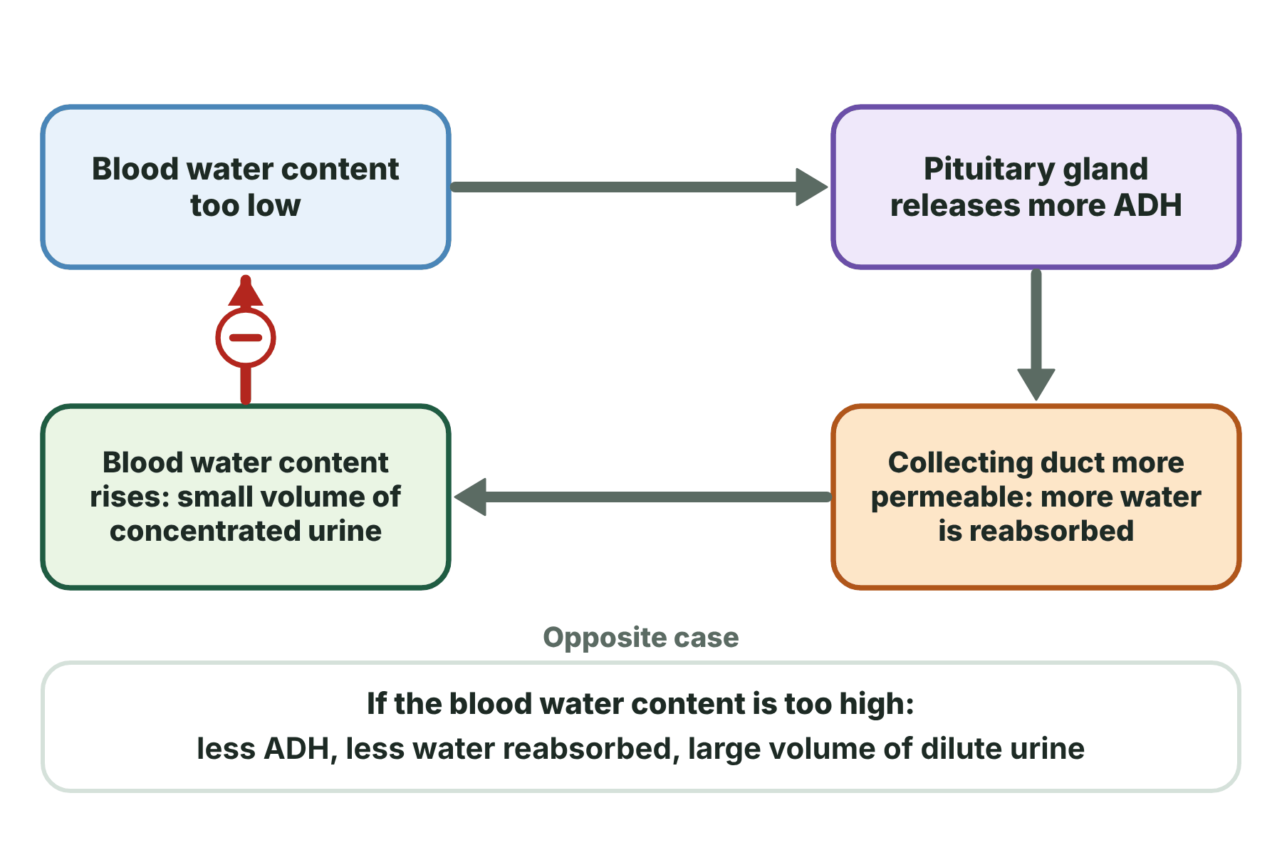
If the blood is too concentrated the pituitary releases more ADH. The collecting duct is more permeable, so more water is reabsorbed and the blood becomes less concentrated. If the blood is too dilute less ADH is released (Higher tier).