Biology · The Nervous System
Scanning and Treating the Brain (Higher tier)
How do doctors see inside a living brain, and why is it so hard to repair? This lesson is for separate Biology only, and for the Higher tier only: Combined Science and Foundation students do not need it. It covers CT and PET scanning and the limits of treating the brain and spinal cord.
Learning Objectives
- 1(Higher tier) Explain why it is difficult to study the brain tissue inside the skull.
- 2(Higher tier) Describe how CT scans and PET scans are used to investigate the structure and the function of the brain.
- 3(Higher tier) Compare the information that CT and PET scans give.
- 4(Higher tier) Explain the limitations in treating damage and disease in the brain and the nervous system, including spinal injuries and brain tumours.
Retrieval practice
-
1
Name the three main regions of the brain.
Show answerHide answer
The cerebral hemispheres, the cerebellum and the medulla oblongata.
-
2
What does the cerebellum do?
Show answerHide answer
Coordinates movement and balance.
-
3
What carries impulses between the brain and the body?
Show answerHide answer
The spinal cord and the nerves.
-
4
What is respiration?
Show answerHide answer
A chemical reaction in cells that releases energy from glucose.
The problem of the inaccessible brain
Doctors have to understand how the brain works so that they can diagnose and treat conditions such as tumours, strokes and dementia. The difficulty is that the brain is very well protected. It is encased in the skull, wrapped in membranes, and made of soft, delicate tissue that is easily damaged. Cutting open the skull to look at a living brain is dangerous and would be a last resort. For this reason, scientists use scanning techniques that take pictures of the inside of the head without opening it.
Why the brain is hard to investigate
There are several reasons why it is difficult to study brain tissue directly.
-
Protected by the skull
The hard bone makes the brain inaccessible, and surgery to expose it carries risk.
-
Delicate tissue
The brain is soft and easily damaged, so taking a sample of living tissue can harm the patient.
-
Hard to see activity
Neurones are very small, and thousands work together, so looking at the tissue cannot show which parts are active.
-
Scans solve the problem
Scans let doctors see the brain, its structure and its activity, from outside the body without any surgery.
CT scanning
A computerised tomography (CT) scan shows the structure of the brain.
-
How it works
An X-ray source and detectors rotate around the head. The X-rays are taken from many angles, and a computer combines them into a series of detailed slices, or a 3D image.
-
What it shows
Different tissues absorb different amounts of X-rays, so bone, brain tissue and fluid show up in different shades. It can show tumours, bleeding, damage after a stroke and injuries to the skull.
-
Advantages
It is fast, so it is used in emergencies such as head injuries and stroke. It is painless and does not involve surgery.
-
Limitations
It shows structure but not activity, and it uses X-rays, which are a form of ionising radiation, so the number of scans has to be limited.
PET scanning
A positron emission tomography (PET) scan shows the function of the brain, which means which parts are active.
-
How it works
The patient is given an injection of glucose that has been made slightly radioactive, called a tracer. Active neurones use more glucose, so more tracer collects in the busy parts of the brain.
-
Detecting the radiation
The tracer gives out radiation, which is picked up by detectors around the head. A computer turns this into a coloured map of the brain, where bright areas are the most active.
-
What it shows
Which areas are active when a person does a task, such as speaking or listening to music. It also shows how the brain differs in diseases such as Alzheimer's disease, or where a tumour is growing quickly.
-
Limitations
The patient is exposed to radiation from the tracer. It is expensive, and the picture is less detailed than a CT scan, so it is often used together with one.
CT and PET scanning
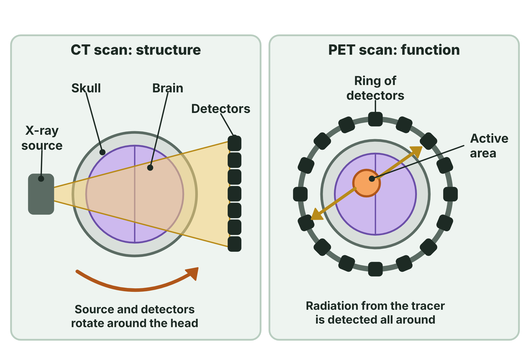
CT scans show the structure of the brain with X-rays. PET scans show active regions by detecting radiation from a tracer.
CT scans and PET scans
CT scan
- Uses X-rays
- Shows the structure of the brain
- Finds tumours, bleeds and damaged areas
- Quick, so it is used in emergencies
PET scan
- Uses a radioactive tracer
- Shows the function of the brain
- Finds which areas are active, and how a disease changes the activity
- Takes longer and is more expensive
Why the brain and nervous system are hard to treat
Even when a problem has been found, repairing the nervous system is very difficult.
-
Neurones do not regrow well
In the brain and spinal cord, neurones that die or are cut are not replaced, so the damage is usually permanent.
-
Delicate and complex
A very small area contains neurones with many different functions, so surgery on one area can damage another by mistake.
-
Many drugs cannot reach the brain
A barrier between the blood and the brain keeps out many chemicals, which protects the brain but also blocks many medicines.
-
Not fully understood
We still do not know exactly how many of the brain's functions work, so it is hard to design treatments.
Spinal injuries
The spinal cord carries all the messages between the brain and the body below the injury.
-
What happens
If the cord is cut or crushed, impulses cannot pass the damaged part, so there is paralysis and loss of feeling below it.
-
Why it is hard to repair
The neurones in the spinal cord do not regenerate, and scar tissue forms at the injury, which blocks the regrowth of axons.
-
Current treatments
Treatment aims to prevent further damage and to help the patient to adapt, using surgery to stabilise the spine, drugs and physiotherapy. Research into stem cells and implants is continuing.
Brain tumours
A tumour in the brain causes problems because of its position and its growth.
-
Pressure on the brain
A growing tumour takes up space inside the skull and presses on brain tissue, which causes headaches and affects the functions of that area.
-
Difficult to remove
Surgery must remove the tumour without damaging healthy tissue, and many tumours lie near vital areas, such as those that control speech or movement.
-
Other treatments are limited
Radiotherapy and chemotherapy can harm healthy tissue, and many drugs cannot cross into the brain.
-
Scans help
CT and PET scans show where the tumour is, how big it is and whether it is growing, which helps surgeons to plan.
Case study
Using scans to find the source of speech
Researchers can ask volunteers to speak or to listen to words while they lie in a PET scanner. Areas of the cerebral hemispheres that are working hard use more glucose, so the tracer builds up there and they show as bright patches. By comparing scans taken during speech and at rest, scientists can map which regions are used for language. The same method shows how the pattern changes in people who have had a stroke.
Summary and exam focus
- The brain is difficult to study because it is protected by the skull and has delicate tissue.
- A CT scan uses X-rays to show the structure of the brain.
- A PET scan uses a radioactive tracer to show the function of the brain, which means the active areas.
- Neurones in the brain and spinal cord do not regenerate well, so damage is often permanent.
- Brain tumours and spinal injuries are hard to treat because of the delicate tissue, the position and the barriers to drugs.
Exam focus
Doctors use CT and PET scans to investigate a patient who is losing the ability to speak. Explain what each type of scan can show, and explain why it is difficult to treat brain damage. (6 marks) (6 marks)
Say that a CT scan shows structure and a PET scan shows function, and link each to the patient, for example a tumour or damage on a CT scan and reduced activity on a PET scan. For treatment, give two reasons from neurones not regenerating, the delicate tissue and drugs not reaching the brain.
Key terms
The words this lesson expects you to use. Each one is linked from the first place it appears above.
- CT scan
- A scan that uses X-rays taken from many angles to make detailed images of the structure of the brain.
- PET scan
- A scan that uses a radioactive tracer to show which areas of the brain are active.
- Tracer
- A radioactive substance injected into the body so that its movement can be detected.
- Radiation
- Energy that travels as waves or particles, such as X-rays or gamma rays.
- Tumour
- An abnormal growth of cells, in this case inside the brain.
- Paralysis
- The loss of the ability to move part of the body.
- Regenerate
- To regrow or replace damaged tissue.
- Spinal injury
- Damage to the spinal cord, which can stop messages passing between the brain and the body.
- Structure
- How something is built and arranged.
- Function
- What something does, or how it works.
You've finished the notes
Check your understanding
Test yourself while it is fresh. Start with the flashcards, then try the exam questions.
Something here looks wrong?
Tell us what and we will go and look. It goes to whoever writes these pages, nobody else, and we do not ask who you are — so there is nothing to sign and nothing comes back to you.