Biology · The Nervous System
The Eye
The eye is a sensory organ full of receptors. This lesson is for separate Biology only: Combined Science students do not need it. It covers how the cornea, lens, iris and retina work together to form an image, and how rods and cones detect light.
Learning Objectives
- 1Describe the structure of the eye and the function of the cornea, lens, iris, retina and optic nerve.
- 2Explain how the iris controls the amount of light entering the eye.
- 3Compare the functions of rod cells and cone cells in the retina.
- 4Describe how the lens changes shape to focus on near and distant objects.
Retrieval practice
-
1
What is a receptor?
Show answerHide answer
A cell that detects a stimulus and turns it into an electrical impulse.
-
2
Which type of receptor is found in the retina?
Show answerHide answer
Light receptors.
-
3
What is the pupil reflex?
Show answerHide answer
A reflex that changes the size of the pupil to control the amount of light entering the eye.
-
4
What is an effector?
Show answerHide answer
A muscle or a gland.
The eye as a sensory organ
The eye is a sensory organ that contains millions of light receptors. It has to do two things: form a clear, focused image on the light-sensitive layer at the back, and change its sensitivity so that you can see in both bright and dim light. The parts of the eye work together to bend the light, control how much enters and detect it. The receptors then send impulses along the optic nerve to the brain, which builds up the picture that you see.
Parts of the eye and what they do
Know where each part is and what job it does.
-
Cornea
The clear, curved layer at the front. It bends (refracts) the light as it enters the eye, and does most of the focusing.
-
Iris
The coloured ring of muscle. It controls the size of the pupil, and so the amount of light entering the eye.
-
Pupil
The hole in the middle of the iris through which the light enters. It looks black.
-
Lens
A clear, flexible structure that bends the light to focus it exactly on the retina. It can change shape to focus on near and distant objects.
-
Retina
The layer at the back that contains the light receptors: rod cells and cone cells. It is where the image is formed.
-
Optic nerve
A bundle of sensory neurones that carries impulses from the retina to the brain.
Structure of the eye
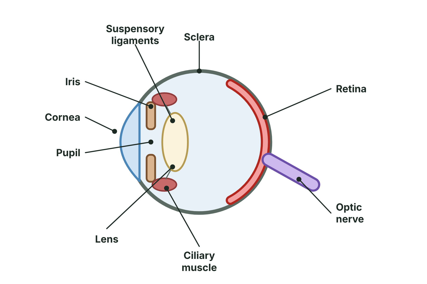
Light enters through the cornea and pupil, is focused by the lens, and is detected by the retina.
How we see an object
Follow the light from the object to the brain.
-
1
Light enters
Light from the object is refracted by the cornea as it enters the eye.
-
2
Through the pupil
The light passes through the pupil. The iris controls how much light gets in.
-
3
Focused by the lens
The lens refracts the light further, so that it is focused into a sharp image on the retina.
-
4
Detected by the retina
The rod and cone cells detect the light and turn it into electrical impulses.
-
5
Impulses to the brain
The impulses travel along the optic nerve, and the brain interprets them as the picture that you see.
The iris and the pupil reflex
The iris protects the retina and helps you see in different light levels. It has two sets of muscle fibres: circular and radial.
-
In bright light
Circular muscles contract and radial muscles relax. The pupil gets smaller, so less light enters the eye and the retina is not damaged.
-
In dim light
Radial muscles contract and circular muscles relax. The pupil gets bigger, so more light enters and you can see better.
-
It is a reflex
The change happens automatically. The receptors in the retina detect the light, and the impulse goes through the brain to the muscles of the iris, which are the effectors.
The pupil in bright and dim light
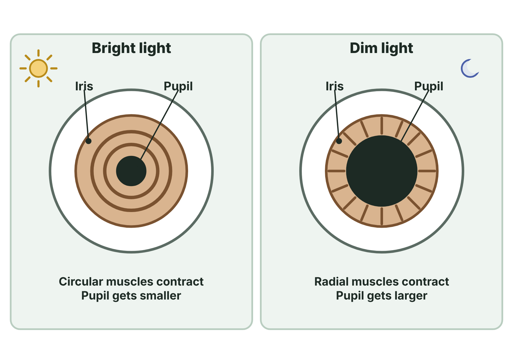
The iris muscles change the size of the pupil, which controls how much light enters the eye.
Rod cells and cone cells
The retina contains two types of light receptor. Together they let us see in dim light and see colour.
-
Rod cells
Very sensitive to light, so they work in dim light. They only give black, white and grey pictures. There are many more rods than cones, mostly towards the edges of the retina.
-
Cone cells
Less sensitive, so they only work in bright light, but they give colour vision. There are three types, sensitive to red, green and blue light. They are most densely packed in the centre of the retina, in the fovea.
-
Why night vision is in black and white
In very dim light only the rods work, so you see in shades of grey, with little colour.
-
Seeing in detail
The fovea has the most cones, so it gives the sharpest, most detailed image, which is why you move your eyes to look straight at something.
Rods and cones
Rod cells
- Work in dim light
- Do not detect colour, so give a black and white image
- Very many of them, mostly around the edge of the retina
- Very sensitive to light
Cone cells
- Work in bright light
- Detect colour, in three types: red, green and blue
- Fewer of them, packed in the fovea at the centre
- Give a sharp, detailed image
Focusing on near and distant objects
The lens changes shape to bring objects at different distances into focus. This is called accommodation. Edexcel only requires the role of the lens, but this is how it does the job.
-
Looking at a near object
The ciliary muscles contract, so the suspensory ligaments become slack. The lens is no longer pulled flat and becomes thicker and more curved, so it bends the light more strongly.
-
Looking at a distant object
The ciliary muscles relax, so the suspensory ligaments are pulled tight. The lens is pulled thinner and flatter, so it bends the light less.
-
Why it matters
Light from near objects spreads out more and needs more bending than light from distant ones. Changing the shape of the lens focuses both on the retina.
Summary and exam focus
- The cornea and lens refract light to focus an image on the retina.
- The iris controls the size of the pupil, and so the amount of light that enters the eye.
- The retina contains rod cells, for dim light, and cone cells, for colour in bright light.
- In bright light the pupil gets smaller, and in dim light it gets bigger.
- The optic nerve carries impulses from the retina to the brain.
Exam focus
Describe the functions of the cornea, the lens, the iris and the retina in the eye, and explain how the iris responds to bright light. (6 marks) (6 marks)
Name each part and give its function in a short phrase using refract, focus, controls the amount of light and detects light. For the iris, include which muscles contract and what happens to the size of the pupil.
Key terms
The words this lesson expects you to use. Each one is linked from the first place it appears above.
- Cornea
- The clear layer at the front of the eye that refracts the light entering it.
- Iris
- The ring of muscle that controls the size of the pupil.
- Pupil
- The hole in the middle of the iris through which light enters the eye.
- Lens
- A flexible, transparent structure that focuses light onto the retina.
- Retina
- The layer at the back of the eye that contains the light receptors.
- Optic nerve
- The nerve that carries impulses from the retina to the brain.
- Rod cell
- A light receptor in the retina that is sensitive in dim light but cannot detect colour.
- Cone cell
- A light receptor in the retina that detects colour in bright light.
- Refraction
- The bending of light as it passes from one transparent substance into another.
- Fovea
- The central part of the retina, packed with cone cells, which gives the sharpest vision.
- Accommodation
- The change in the shape of the lens to focus on near or distant objects.
You've finished the notes
Check your understanding
Test yourself while it is fresh. Start with the flashcards, then try the exam questions.
Something here looks wrong?
Tell us what and we will go and look. It goes to whoever writes these pages, nobody else, and we do not ask who you are — so there is nothing to sign and nothing comes back to you.