Exam questions · Biology
Exchange and Transport in Animals
- 36 exam questions
- 96 marks
- 40 quick checks
Surface Area to Volume Ratio and Exchange Surfaces
Just this lesson-
1 State [1 mark]
State what is meant by surface area to volume ratio.
Show answerHide answer
Model answer
The surface area of an object divided by its volume.
Mark scheme
- Surface area divided by volume — 1 mark
-
2 Calculate [2 marks]
A cube has sides of 2 cm. Calculate its surface area to volume ratio.
Show answerHide answer
Model answer
Surface area = 6 × 2 × 2 = 24 cm². Volume = 2 × 2 × 2 = 8 cm³. Ratio = 24 ÷ 8 = 3, so 3:1.
Mark scheme
- Surface area 24 cm² and volume 8 cm³ — 1 mark
- Ratio of 3:1 — 1 mark
-
3 Calculate [3 marks]
Cube A has sides of 1 cm. Cube B has sides of 3 cm. Calculate the surface area to volume ratio of each cube, and state which has the larger ratio.
Show answerHide answer
Model answer
Cube A: 6 ÷ 1 = 6, so 6:1. Cube B: 54 ÷ 27 = 2, so 2:1. Cube A, the smaller cube, has the larger ratio.
Mark scheme
- Ratio of 6:1 for cube A — 1 mark
- Ratio of 2:1 for cube B — 1 mark
- Cube A has the larger ratio — 1 mark
-
4 Explain [2 marks]
Explain why a single-celled organism does not need a transport system.
Show answerHide answer
Model answer
It has a large surface area to volume ratio. So diffusion across its cell membrane is fast enough to supply the whole cell.
Mark scheme
- Large surface area to volume ratio — 1 mark
- Diffusion across the surface is fast enough / short distances — 1 mark
-
5 Explain [3 marks]
Explain why a large multicellular organism needs exchange surfaces and a transport system.
Show answerHide answer
Model answer
A large organism has a small surface area to volume ratio, so its body surface is too small to supply all its cells. Cells deep inside are far from the outside, so diffusion over that distance would be too slow. Exchange surfaces swap materials quickly and a transport system carries them to every cell.
Mark scheme
- Small surface area to volume ratio — 1 mark
- Diffusion distances too long / too slow — 1 mark
- Exchange surface takes in materials and the transport system carries them to the cells — 1 mark
-
6 Name [3 marks]
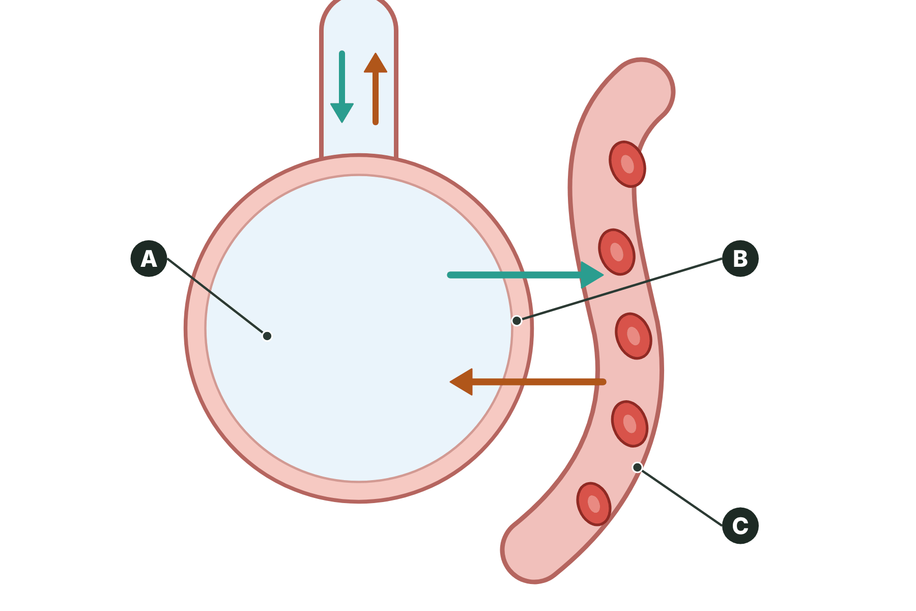
Figure 1 shows a gas exchange surface in the lungs. Name the parts labelled A, B and C.
Show answerHide answer
Model answer
A is an alveolus (air sac). B is the thin wall of the alveolus (one cell thick). C is a capillary.
Mark scheme
- A: alveolus — 1 mark
- B: wall (of the alveolus) — 1 mark
- C: capillary — 1 mark
-
7 Explain [4 marks]
Explain how the small intestine is adapted for the absorption of digested food.
Show answerHide answer
Model answer
The inner wall is covered with villi, which give a large surface area. Each villus has a thin wall, so the diffusion path is short. Each villus has a rich blood supply, which carries the absorbed food away and keeps the concentration gradient steep. This allows food to be absorbed quickly.
Mark scheme
- Villi give a large surface area — 1 mark
- Thin wall, so a short diffusion path — 1 mark
- Rich blood supply — 1 mark
- Carries absorbed food away / keeps a steep concentration gradient — 1 mark
-
8 Explain [6 marks]
Explain how the structure of the lungs and the structure of a fish's gills are adapted for gas exchange. In your answer, refer to surface area to volume ratio.
Show answerHide answer
Model answer
Large organisms have a small surface area to volume ratio, so they need specialised gas exchange surfaces. The lungs contain millions of alveoli, which give a very large surface area. Each alveolus has a wall one cell thick, so the diffusion path is short, and is surrounded by capillaries, so the blood supply is rich. Breathing ventilates the lungs, so oxygen is always at a higher concentration in the alveoli than in the blood. In a fish, the gills are made of many thin filaments, which give a large surface area. The filaments have thin surfaces and a rich blood supply, and water flowing over them maintains the concentration gradient.
Mark scheme
- Small surface area to volume ratio means a specialised surface is needed — 1 mark
- Alveoli / gill filaments give a large surface area — 1 mark
- Thin walls, short diffusion path — 1 mark
- Rich blood supply in both — 1 mark
- Ventilation of the lungs / water flowing over the gills — 1 mark
- Keeps a steep concentration gradient — 1 mark
Quick check
-
1
What is the surface area to volume ratio of a cube with sides of 2 cm?
Show answerHide answer
C: 3:1
Surface area is 6 × 4 = 24 cm² and volume is 8 cm³, so the ratio is 24 ÷ 8 = 3:1.
-
2
What happens to the surface area to volume ratio of an organism as it gets bigger?
Show answerHide answer
B: It gets smaller
Volume grows faster than surface area, so the ratio gets smaller.
-
3
Why does a large animal need a transport system?
Show answerHide answer
D: Diffusion over long distances is too slow
Its surface area to volume ratio is small, so diffusion alone is too slow to supply cells deep inside.
-
4
Which feature of an exchange surface keeps the concentration gradient steep in an animal?
Show answerHide answer
A: An efficient blood supply
The blood carries substances away from the exchange surface, so the concentration there stays low and the gradient stays steep.
-
5
What are the tiny air sacs in the lungs called?
Show answerHide answer
C: Alveoli
Alveoli are the tiny air sacs where gas exchange happens.
-
6
How do villi help the small intestine absorb food?
Show answerHide answer
B: They increase the surface area
Villi are finger-like folds, so they greatly increase the surface area for absorption.
-
7
Which of these is an exchange surface in a plant?
Show answerHide answer
D: A root hair cell
Root hair cells have long thin extensions that give a large surface area for taking up water and ions.
-
8
A cube is made 3 times wider. What happens to its surface area to volume ratio?
Show answerHide answer
B: It falls to a third
A 1 cm cube has a ratio of 6:1 and a 3 cm cube has 2:1, so the ratio falls to a third.
The Heart and Double Circulation
Just this lesson-
1 State [1 mark]
Name the blood vessel that carries blood from the heart to the lungs.
Show answerHide answer
Model answer
The pulmonary artery.
Mark scheme
- Pulmonary artery — 1 mark
-
2 Name [3 marks]
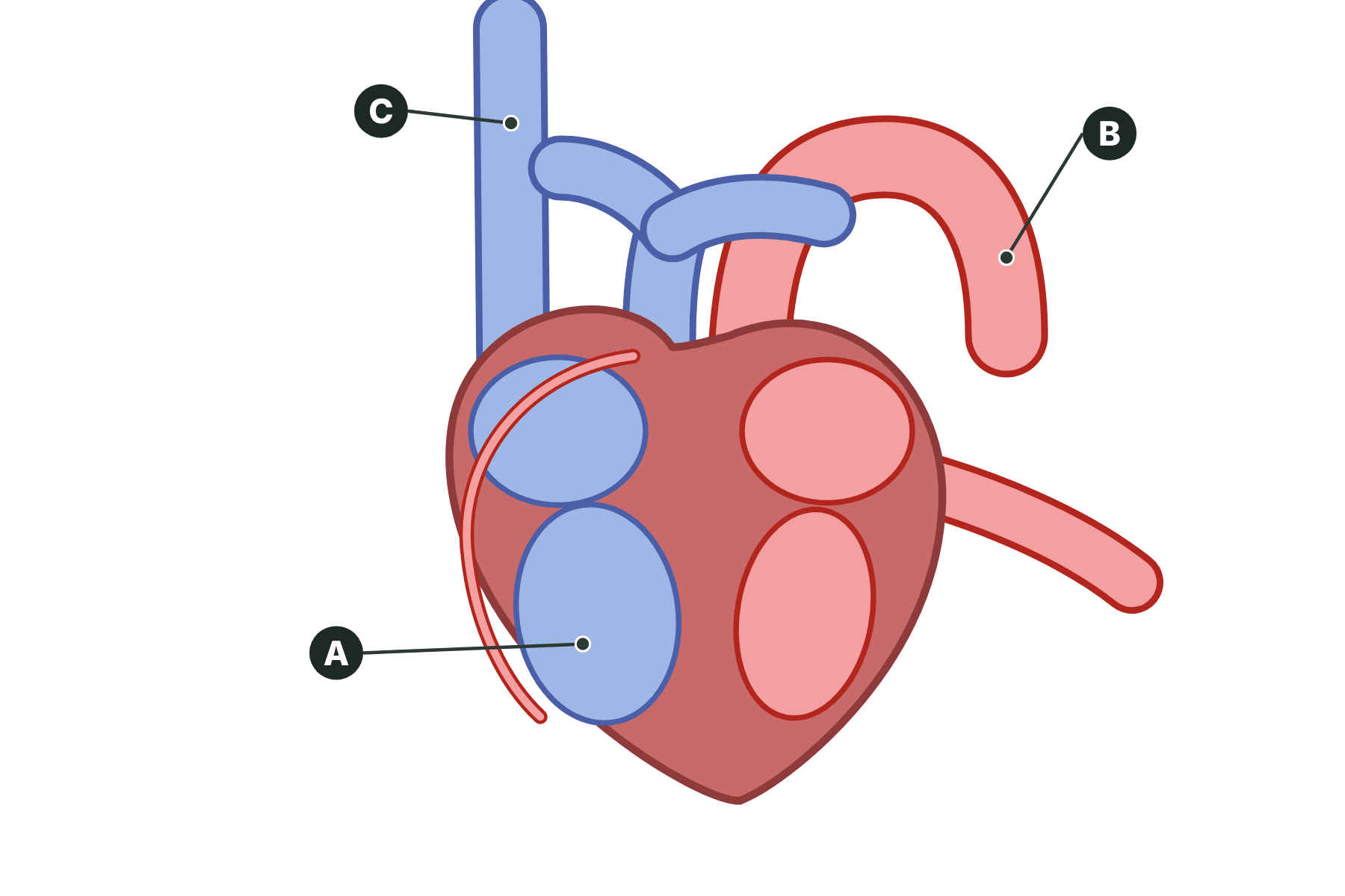
Figure 1 shows the human heart. Name the parts labelled A, B and C.
Show answerHide answer
Model answer
A is the right ventricle. B is the aorta. C is the vena cava.
Mark scheme
- A: right ventricle — 1 mark
- B: aorta — 1 mark
- C: vena cava — 1 mark
-
3 Describe [2 marks]
Describe the function of the coronary arteries.
Show answerHide answer
Model answer
They supply the heart muscle with oxygenated blood. This gives the muscle the oxygen it needs for respiration to release energy for contraction.
Mark scheme
- Supply the heart (muscle) with blood — 1 mark
- Oxygen for respiration — 1 mark
-
4 Explain [2 marks]
Explain why the wall of the left ventricle is thicker than the wall of the right ventricle.
Show answerHide answer
Model answer
The left ventricle pumps blood around the whole body, which is a long way, so it needs to generate a higher pressure. A thicker muscle wall contracts with more force.
Mark scheme
- Pumps blood to the whole body / further — 1 mark
- Thicker muscle gives a greater force / higher pressure — 1 mark
-
5 Calculate [2 marks]
A student counts 18 pulses in 15 seconds. Calculate the heart rate in beats per minute.
Show answerHide answer
Model answer
There are four 15-second periods in a minute. 18 × 4 = 72 beats per minute.
Mark scheme
- Multiplies by 4 — 1 mark
- 72 beats per minute — 1 mark
-
6 Explain [3 marks]
Explain why humans have a double circulatory system.
Show answerHide answer
Model answer
Blood loses pressure as it passes through the capillaries in the lungs. Returning it to the heart increases the pressure again before it is sent to the body. This means the blood reaches the body quickly, while the pressure in the lungs can stay low enough not to damage the thin alveoli.
Mark scheme
- Blood loses pressure in the lung capillaries — 1 mark
- The heart increases the pressure again — 1 mark
- Low pressure in the lungs protects them / higher pressure for the body — 1 mark
-
7 Describe [4 marks]
Describe how the natural pacemaker and an artificial pacemaker control the heart rate.
Show answerHide answer
Model answer
The natural pacemaker is a group of cells in the right atrium. These cells send out electrical signals that make the heart muscle contract, which sets the natural resting heart rate. An artificial pacemaker is an electrical device. It sends regular electrical pulses to the heart to correct an irregular heartbeat.
Mark scheme
- Natural pacemaker in the right atrium — 1 mark
- Cells send electrical signals that make the heart contract — 1 mark
- Artificial pacemaker is an electrical device — 1 mark
- It corrects irregularities in the heart rate — 1 mark
Quick check
-
1
Which chamber of the heart pumps blood to the body?
Show answerHide answer
D: Left ventricle
The left ventricle pumps oxygenated blood into the aorta, which carries it round the body.
-
2
Which vessel brings oxygenated blood from the lungs to the heart?
Show answerHide answer
B: Pulmonary vein
The pulmonary vein carries oxygenated blood from the lungs to the left atrium.
-
3
What do the coronary arteries supply?
Show answerHide answer
C: The heart muscle with blood
The coronary arteries run over the surface of the heart and bring oxygenated blood to the heart muscle.
-
4
Why is the wall of the left ventricle thicker than that of the right?
Show answerHide answer
A: It pumps blood further
It has to pump blood to the whole body, so it needs to contract with more force.
-
5
Where is the natural pacemaker of the heart?
Show answerHide answer
D: The right atrium
A group of cells in the right atrium sets the natural resting heart rate.
-
6
A pulse is counted as 20 beats in 15 seconds. What is the heart rate?
Show answerHide answer
C: 80 beats per minute
20 × 4 = 80 beats per minute.
-
7
Why do humans have a double circulatory system?
Show answerHide answer
B: To keep the pressure high round the body
The blood loses pressure in the lungs, so it returns to the heart to be pumped again to the body.
-
8
What does an artificial pacemaker do?
Show answerHide answer
A: Corrects an irregular heart rate
It sends electrical pulses to correct an irregular heartbeat.
Blood Vessels
Just this lesson-
1 State [1 mark]
Name the type of blood vessel that carries blood away from the heart.
Show answerHide answer
Model answer
Artery.
Mark scheme
- Artery — 1 mark
-
2 Name [3 marks]
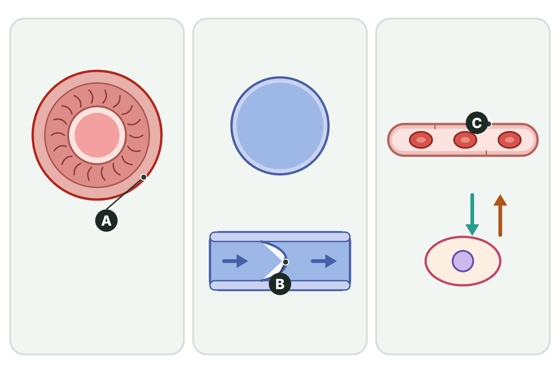
Figure 1 shows three types of blood vessel. Name the parts labelled A, B and C.
Show answerHide answer
Model answer
A is the (muscular) wall of the artery. B is a valve. C is the wall of the capillary.
Mark scheme
- A: wall of the artery / muscle and elastic fibres — 1 mark
- B: valve — 1 mark
- C: wall of the capillary — 1 mark
-
3 Explain [2 marks]
Explain why veins have valves.
Show answerHide answer
Model answer
The blood in veins is at low pressure, so it could flow backwards. The valves close to stop this happening, so the blood flows only towards the heart.
Mark scheme
- Blood at low pressure could flow backwards — 1 mark
- Valves prevent backflow — 1 mark
-
4 Explain [3 marks]
Explain how the structure of an artery is related to its function.
Show answerHide answer
Model answer
An artery carries blood away from the heart at high pressure. Its wall is thick, with muscle and elastic fibres, so that it can withstand the pressure. The elastic fibres let the wall stretch and recoil as blood surges through.
Mark scheme
- Carries blood at high pressure — 1 mark
- Thick wall with muscle and elastic fibres — 1 mark
- Withstands the pressure / stretches and recoils — 1 mark
-
5 Explain [3 marks]
Explain how a capillary is adapted for exchanging substances with the cells.
Show answerHide answer
Model answer
The wall of a capillary is only one cell thick, so the diffusion path is short. Capillaries form huge networks, which give a large surface area. The blood moves slowly through them, which gives time for substances to diffuse.
Mark scheme
- Wall one cell thick / short diffusion path — 1 mark
- Large surface area from many capillaries — 1 mark
- Slow flow gives time for diffusion — 1 mark
-
6 Calculate [2 marks]
A volume of 450 cm³ of blood flows through a blood vessel in 90 seconds. Calculate the rate of blood flow in cm³ per second.
Show answerHide answer
Model answer
Rate = volume ÷ time = 450 ÷ 90 = 5 cm³ per second.
Mark scheme
- 450 ÷ 90 — 1 mark
- 5 cm³ per second — 1 mark
-
7 Compare [4 marks]
Compare the structure of an artery with the structure of a vein.
Show answerHide answer
Model answer
The wall of an artery is thick, with muscle and elastic fibres, but the wall of a vein is thinner. An artery has a narrow lumen but a vein has a wider lumen. Veins have valves but arteries do not. Arteries carry blood at high pressure away from the heart, but veins carry blood at low pressure to the heart.
Mark scheme
- Artery wall thicker than vein wall — 1 mark
- Artery lumen narrower than vein lumen — 1 mark
- Veins have valves, arteries do not — 1 mark
- Artery high pressure away from the heart, vein low pressure towards it — 1 mark
Quick check
-
1
Which blood vessel has valves?
Show answerHide answer
B: Vein
Veins have valves to stop the low-pressure blood flowing backwards.
-
2
Why is the wall of a capillary only one cell thick?
Show answerHide answer
C: To give a short diffusion path
A thin wall gives a short diffusion path, so substances move across quickly.
-
3
What do arteries carry?
Show answerHide answer
A: Blood away from the heart
Arteries carry blood away from the heart, and this blood is at high pressure.
-
4
Which blood vessel carries deoxygenated blood away from the heart?
Show answerHide answer
D: Pulmonary artery
The pulmonary artery carries deoxygenated blood from the right ventricle to the lungs.
-
5
What gives artery walls the ability to stretch and recoil?
Show answerHide answer
B: Elastic fibres
Elastic fibres in the wall let it stretch with each surge of blood and then recoil.
-
6
900 cm³ of blood flows through a vessel in 3 minutes. What is the rate of blood flow?
Show answerHide answer
C: 300 cm³ per minute
Rate = volume ÷ time = 900 ÷ 3 = 300 cm³ per minute.
-
7
Why does blood flow slowly through capillaries?
Show answerHide answer
A: To give time for exchange
Slow flow gives more time for substances to diffuse between the blood and the cells.
-
8
Which statement about veins is correct?
Show answerHide answer
D: They have a wide lumen and valves
Veins return blood to the heart at low pressure, so they have a wide lumen and valves.
The Lungs and Gas Exchange
Just this lesson-
1 Name [3 marks]
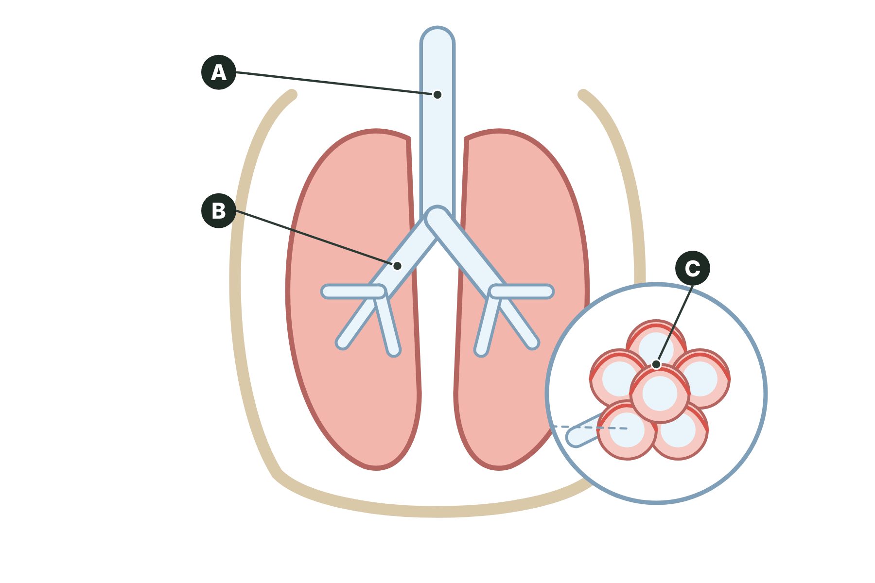
Figure 1 shows the lungs. Name the parts labelled A, B and C.
Show answerHide answer
Model answer
A is the trachea. B is a bronchus. C is the alveoli (air sacs).
Mark scheme
- A: trachea — 1 mark
- B: bronchus — 1 mark
- C: alveoli — 1 mark
-
2 State [2 marks]
Name the two gases exchanged in the alveoli and state the direction each one moves.
Show answerHide answer
Model answer
Oxygen moves from the alveoli into the blood. Carbon dioxide moves from the blood into the alveoli.
Mark scheme
- Oxygen from the alveoli into the blood — 1 mark
- Carbon dioxide from the blood into the alveoli — 1 mark
-
3 Explain [2 marks]
Explain why the walls of the alveoli are only one cell thick.
Show answerHide answer
Model answer
A thin wall gives a short diffusion path. Oxygen and carbon dioxide can therefore diffuse across quickly.
Mark scheme
- Short diffusion path — 1 mark
- Faster diffusion of gases — 1 mark
-
4 Explain [3 marks]
Explain why oxygen diffuses from the alveoli into the blood.
Show answerHide answer
Model answer
The concentration of oxygen is higher in the air in the alveoli than in the blood. Oxygen therefore diffuses down the concentration gradient. The gradient is kept steep by breathing and by the blood carrying oxygen away.
Mark scheme
- Higher concentration of oxygen in the alveoli — 1 mark
- Diffuses down the concentration gradient — 1 mark
- Gradient kept steep by ventilation / blood flow — 1 mark
-
5 Calculate [2 marks]
A person takes 15 breaths per minute. Each breath takes in 400 cm³ of air. Calculate the volume of air breathed in per minute.
Show answerHide answer
Model answer
400 × 15 = 6000 cm³ per minute.
Mark scheme
- 400 × 15 — 1 mark
- 6000 cm³ per minute — 1 mark
-
6 Explain [4 marks]
Explain how the lungs are adapted for gaseous exchange.
Show answerHide answer
Model answer
The lungs contain millions of alveoli, which give a very large surface area. The walls of the alveoli and capillaries are one cell thick, so the diffusion path is short. A rich supply of blood in the capillaries carries oxygen away and brings carbon dioxide, keeping the concentration gradient steep. Breathing ventilates the alveoli, which also keeps the gradient steep.
Mark scheme
- Millions of alveoli give a large surface area — 1 mark
- Walls one cell thick, short diffusion path — 1 mark
- Rich blood supply keeps the gradient steep — 1 mark
- Ventilation keeps the gradient steep — 1 mark
-
7 Explain [3 marks]
A person has emphysema, a disease that destroys the walls of the alveoli. Explain why this person feels short of breath.
Show answerHide answer
Model answer
Destroying the walls reduces the surface area of the alveoli. Less oxygen can diffuse into the blood. The body cells therefore receive less oxygen for respiration, so the person feels short of breath.
Mark scheme
- Reduced surface area — 1 mark
- Less oxygen diffuses into the blood — 1 mark
- Less oxygen for respiration in cells — 1 mark
Quick check
-
1
What are the tiny air sacs in the lungs called?
Show answerHide answer
C: Alveoli
Alveoli are the air sacs where gases are exchanged with the blood.
-
2
Which gas diffuses from the blood into the alveoli?
Show answerHide answer
B: Carbon dioxide
Carbon dioxide is at a higher concentration in the blood arriving at the lungs, so it diffuses into the alveoli.
-
3
What is the trachea?
Show answerHide answer
A: The windpipe
The trachea is the windpipe, which carries air from the mouth and nose to the bronchi.
-
4
How many cells thick is the wall of an alveolus?
Show answerHide answer
D: One
The wall is one cell thick, which gives a short diffusion path.
-
5
Which feature of the lungs keeps the concentration gradients steep by moving air?
Show answerHide answer
C: Ventilation
Ventilation, or breathing, moves fresh air in and stale air out.
-
6
What surrounds each alveolus?
Show answerHide answer
B: A network of capillaries
A network of capillaries brings blood past the alveolus for gas exchange.
-
7
A person breathes 20 times a minute and takes in 300 cm³ each time. What volume do they breathe in per minute?
Show answerHide answer
D: 6000 cm³
300 × 20 = 6000 cm³ per minute.
-
8
Why do the lungs have millions of alveoli?
Show answerHide answer
A: To give a large surface area
Many alveoli give a very large surface area for gas exchange.
Blood
Just this lesson-
1 State [1 mark]
Name the part of the blood that carries oxygen.
Show answerHide answer
Model answer
Red blood cells (haemoglobin in the red blood cells).
Mark scheme
- Red blood cells — 1 mark
-
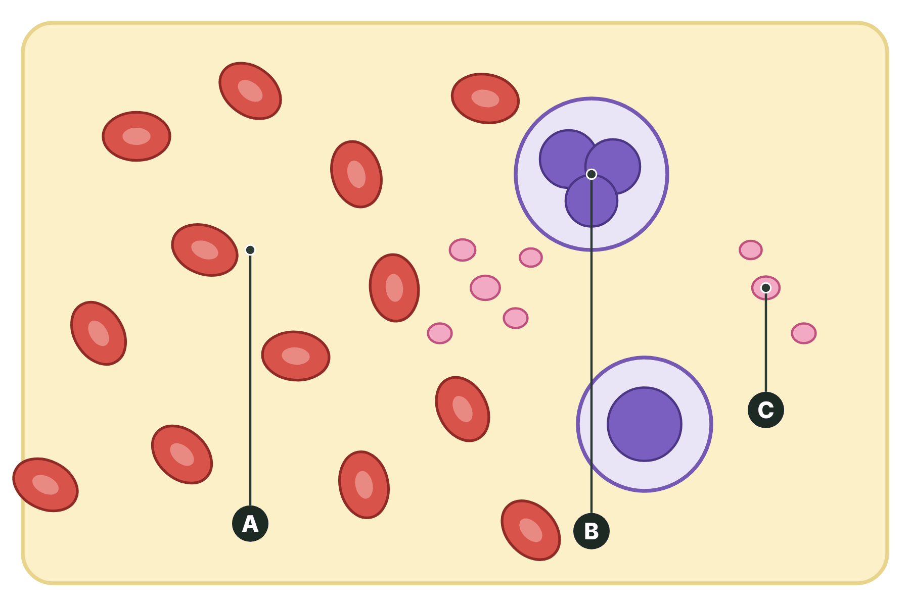
2 Name [3 marks]
Figure 1 shows some of the components of blood. Name the parts labelled A, B and C.
Show answerHide answer
Model answer
A is a red blood cell. B is a white blood cell. C is a platelet.
Mark scheme
- A: red blood cell — 1 mark
- B: white blood cell — 1 mark
- C: platelet — 1 mark
-
3 State [2 marks]
Name two substances carried in the plasma, and state where each one is carried to.
Show answerHide answer
Model answer
Carbon dioxide is carried to the lungs. Urea is carried to the kidneys. Accept glucose carried to the body cells, or hormones carried to their target organs.
Mark scheme
- A named substance — 1 mark
- Its correct destination — 1 mark
-
4 Explain [3 marks]
Explain how a red blood cell is adapted to carry oxygen.
Show answerHide answer
Model answer
It contains haemoglobin, which joins with oxygen. It has no nucleus, so there is more room for haemoglobin. Its biconcave shape gives a large surface area for oxygen to diffuse in and out.
Mark scheme
- Contains haemoglobin — 1 mark
- No nucleus so more room for haemoglobin — 1 mark
- Biconcave shape gives a large surface area — 1 mark
-
5 Describe [2 marks]
Describe the function of platelets.
Show answerHide answer
Model answer
Platelets help the blood to clot at a wound. The clot forms a scab that stops the bleeding and prevents microorganisms entering.
Mark scheme
- Help the blood to clot — 1 mark
- Stops blood loss / stops pathogens getting in — 1 mark
-
6 Describe [3 marks]
A photograph of blood shows many small round cells, a few much larger cells with a dark purple centre, and some tiny fragments. Describe how you can identify each of these using what you know about blood cells.
Show answerHide answer
Model answer
The many small round cells with no nucleus are red blood cells. The few larger cells with a dark stained nucleus are white blood cells. The tiny fragments with no nucleus are platelets.
Mark scheme
- Small round cells with no nucleus are red blood cells — 1 mark
- Larger cells with a nucleus are white blood cells — 1 mark
- Tiny fragments are platelets — 1 mark
-
7 Evaluate [4 marks]
Blood transfusions use blood products from donors. Evaluate the risks and benefits of using blood products.
Show answerHide answer
Model answer
A transfusion can save the life of a person who has lost a lot of blood or who has a blood disorder. However, there are risks. The donor blood could be infected with a pathogen, although screening reduces this risk. If the blood group is not matched, the patient can have a serious reaction. Some patients may have an allergic reaction. On balance the benefits usually outweigh the risks because blood is screened and matched.
Mark scheme
- Benefit: saves life / replaces lost blood — 1 mark
- Risk: infection from donor blood — 1 mark
- Risk: mismatched blood group / allergic reaction — 1 mark
- A conclusion weighing risks and benefits — 1 mark
Quick check
-
1
Which part of the blood carries oxygen?
Show answerHide answer
B: Red blood cells
Red blood cells contain haemoglobin, which joins with oxygen.
-
2
Which part of the blood helps it to clot?
Show answerHide answer
D: Platelets
Platelets are cell fragments that help blood clot at a wound.
-
3
Which blood cell has a nucleus?
Show answerHide answer
C: White blood cell
White blood cells have a nucleus. Red blood cells and platelets do not.
-
4
What is carried in the plasma from the liver to the kidneys?
Show answerHide answer
A: Urea
Urea is made in the liver and carried in the plasma to the kidneys.
-
5
Why do red blood cells have no nucleus?
Show answerHide answer
B: To make more room for haemoglobin
It leaves more room for haemoglobin, so the cell can carry more oxygen.
-
6
What is the red pigment in red blood cells?
Show answerHide answer
C: Haemoglobin
Haemoglobin joins with oxygen in the lungs and releases it in the tissues.
-
7
What do phagocytes do?
Show answerHide answer
D: Engulf pathogens
Phagocytes are white blood cells that engulf and digest pathogens.
-
8
Which component of blood is a fragment of a cell?
Show answerHide answer
A: Platelets
Platelets are small fragments of cells with no nucleus.