Biology · Blood, Blood Vessels and the Heart
The Heart and Double Circulation
The heart is two pumps in one, and the blood passes through it twice on every trip round the body. This lesson is for both Combined Science and separate Biology, at Foundation and Higher. It explains how the structure of the heart and the circulatory system is related to their function, including the major blood vessels, the valves, the thickness of the chamber walls and the double circulation.
Learning Objectives
- 1Describe the structure of the heart: the four chambers, the septum, the valves and the major blood vessels.
- 2Explain how the relative thickness of the walls of the chambers is related to their function.
- 3Describe the route that blood takes through the heart, the lungs and the body, and say whether it is oxygenated at each stage.
- 4Explain why humans have a double circulation.
Retrieval practice
-
1
Which vessels carry blood away from the heart?
Show answerHide answer
Arteries.
-
2
Why do veins have valves?
Show answerHide answer
To stop the blood flowing backwards, because the pressure in veins is low.
-
3
Which blood cells carry oxygen, and in which substance?
Show answerHide answer
Red blood cells, in haemoglobin.
-
4
Which gases are exchanged in the alveoli?
Show answerHide answer
Oxygen diffuses into the blood and carbon dioxide diffuses out.
Two pumps side by side
The heart is a muscular organ, about the size of a fist, made mainly of cardiac muscle that contracts without tiring. It is really two pumps joined together. The right side pumps deoxygenated blood to the lungs, and the left side pumps oxygenated blood to the rest of the body. A wall of muscle, the septum, keeps the two sides apart so that the two kinds of blood do not mix.
The four chambers
Each side of the heart has an upper chamber that receives blood and a lower chamber that pumps it out.
-
Atria
The two upper chambers, the right atrium and the left atrium, receive blood from the veins. Their walls are thin, because they only have to push the blood a short way down into the ventricles.
-
Ventricles
The two lower chambers, the right ventricle and the left ventricle, pump blood out of the heart into the arteries. Their walls are thicker than those of the atria because they have to push blood much further.
-
The septum
The septum is the wall of muscle between the left and right sides. It stops oxygenated blood on the left from mixing with deoxygenated blood on the right.
-
Thickness of the walls
The left ventricle has the thickest wall because it has to pump blood at high pressure all the way round the body. The right ventricle has a thinner wall because it only pumps blood to the nearby lungs, where a lower pressure is enough and the delicate capillaries would be damaged by a high one.
The major blood vessels
Four large vessels carry blood into and out of the heart. Remember that arteries carry blood away from the heart and veins carry it towards the heart.
-
Vena cava
A large vein that brings deoxygenated blood from the body into the right atrium.
-
Pulmonary artery
Carries deoxygenated blood from the right ventricle to the lungs. It is the only artery that carries deoxygenated blood.
-
Pulmonary vein
Brings oxygenated blood from the lungs into the left atrium. It is the only vein that carries oxygenated blood.
-
Aorta
The largest artery. It carries oxygenated blood from the left ventricle to the body. The coronary arteries branch off it to supply the heart muscle itself with blood.
The valves
Valves make sure that the blood flows one way through the heart. They are opened by blood pushing from behind and closed by blood pushing back.
-
Atrioventricular valves
These are between each atrium and ventricle. They open when the atrium contracts, so blood flows down into the ventricle. They close when the ventricle contracts, so the blood cannot flow back into the atrium. Tendons attach them to the ventricle wall so that they are not pushed inside out.
-
Semilunar valves
These are at the start of the pulmonary artery and the aorta. They open when the ventricle contracts and blood is forced out, and close when the ventricle relaxes so that blood cannot fall back into the ventricle.
-
The sound of the heart
The heart sounds, the lub-dub of a heartbeat, are made by the valves closing.
-
One-way flow
The valves make the heart an efficient pump, because none of the blood pushed out can return.
Inside the heart
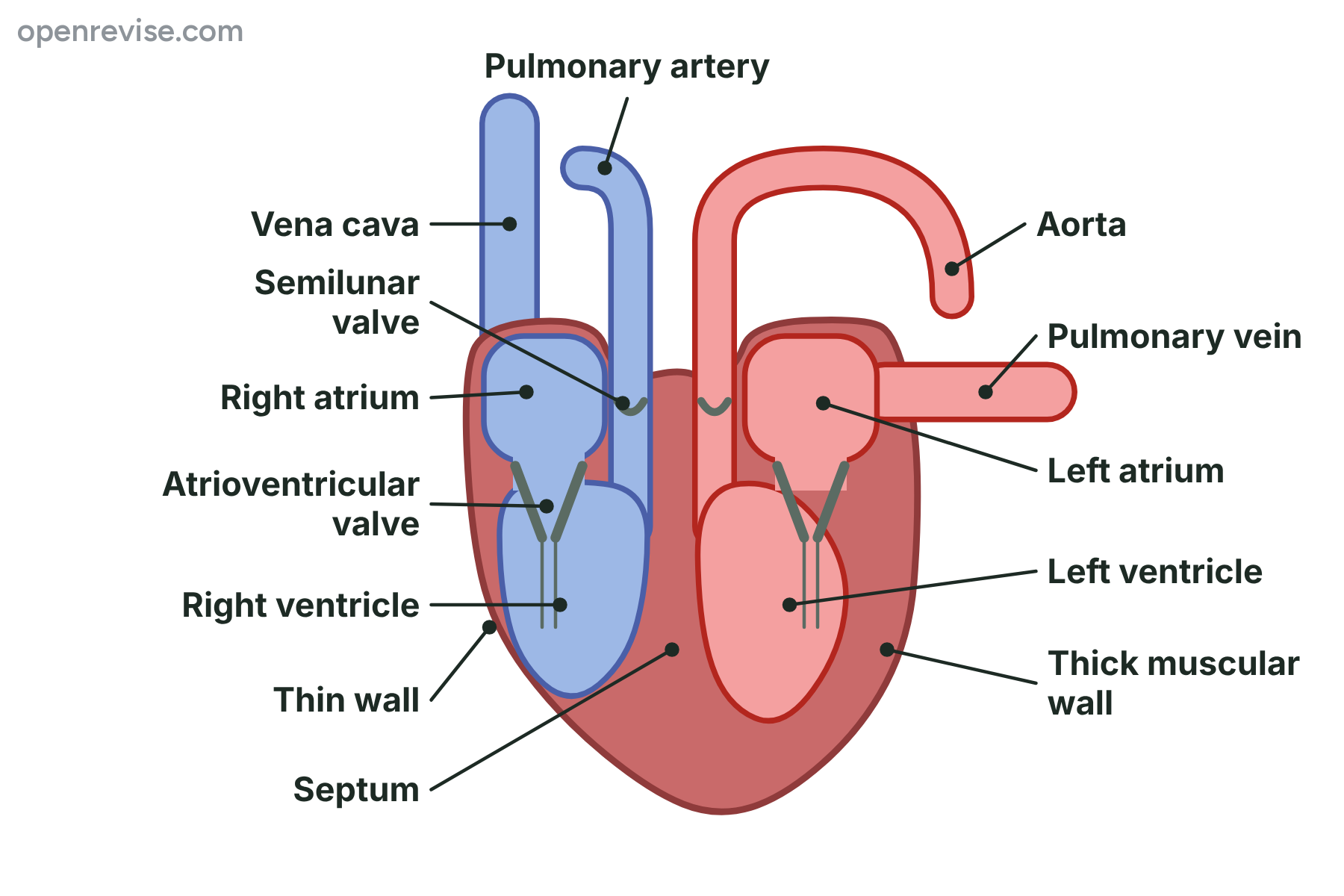
The left ventricle has the thickest wall because it pumps blood all the way round the body.
The right side and the left side of the heart
Right side
- Receives deoxygenated blood from the body through the vena cava
- Pumps the blood to the lungs through the pulmonary artery
- The right ventricle has a thinner wall
- A lower pressure is enough
Left side
- Receives oxygenated blood from the lungs through the pulmonary vein
- Pumps the blood to the body through the aorta
- The left ventricle has a much thicker wall
- A high pressure is needed
The route of the blood
Follow one drop of blood from the body, through the heart and lungs and back round the body.
-
1
Vena cava
Deoxygenated blood from the body flows in through the vena cava.
-
2
Right atrium to right ventricle
The right atrium contracts, and the blood passes through the atrioventricular valve into the right ventricle.
-
3
To the lungs
The right ventricle contracts and forces the blood through the semilunar valve into the pulmonary artery and on to the lungs.
-
4
Gas exchange
In the lungs the blood loses carbon dioxide and picks up oxygen.
-
5
Pulmonary vein to left atrium
Oxygenated blood returns in the pulmonary vein to the left atrium.
-
6
Left atrium to left ventricle
The left atrium contracts, and the blood passes through the atrioventricular valve into the left ventricle.
-
7
To the body
The left ventricle contracts strongly and forces the blood through the semilunar valve into the aorta and out to the body.
-
8
Back again
The blood gives up oxygen to the cells and takes in carbon dioxide, then returns to the vena cava.
The double circulation
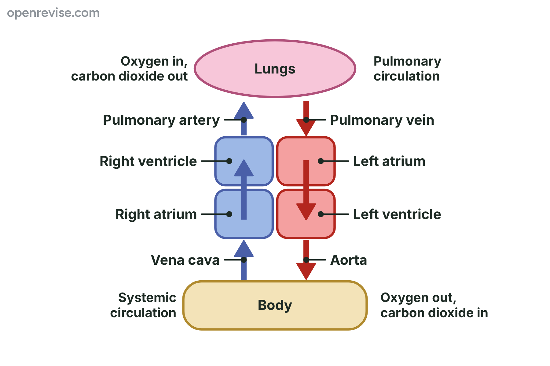
The blood passes through the heart twice on each complete trip: once to the lungs and once to the body.
Why humans have a double circulation
The circulatory system is called a double circulation because it has two circuits that are joined at the heart.
-
Two circuits
The pulmonary circulation takes blood from the heart to the lungs and back. The systemic circulation takes blood from the heart to the body and back. Blood passes through the heart twice on each complete trip.
-
Pressure is restored
The blood loses pressure as it squeezes through the tiny capillaries in the lungs. Returning it to the heart lets the left ventricle pump it again at high pressure, so it reaches the body quickly.
-
Blood is kept separate
The septum keeps oxygenated and deoxygenated blood apart, so the blood sent to the body is fully loaded with oxygen. This suits active animals that need a lot of oxygen.
-
The lungs are protected
The right ventricle pumps blood to the lungs at a lower pressure, so the thin walls of the alveoli and capillaries are not damaged.
Single circulation and double circulation
Fish: single circulation
- The heart has two chambers
- Blood passes through the heart once on each trip
- The blood goes from the heart to the gills and then straight to the body
- The pressure drops after the gills, so the blood flows slowly to the body
Mammals: double circulation
- The heart has four chambers
- Blood passes through the heart twice on each trip
- The blood returns to the heart after the lungs
- The pressure is raised again, so oxygen reaches the body quickly
Comparing the thickness of the walls
A typical left ventricle wall is 12 mm thick and a typical right ventricle wall is 4 mm thick. Calculate how many times thicker the left ventricle wall is, and say why.
Show the solutionHide the solution
- 1 Divide the thicknesses \(12 \div 4 = 3\)
- 2 State the comparison The left ventricle wall is 3 times as thick as the right
- 3 Explain The left ventricle pushes blood to the whole body, so it needs more muscle to generate a higher pressure. The right ventricle only pushes blood to the lungs
AnswerThe left ventricle wall is 3 times thicker, because it pumps blood at a higher pressure all round the body.
Case study
Feeding the heart muscle
The heart muscle needs its own supply of oxygen and glucose. It gets them from the coronary arteries, which branch off the aorta and spread over the surface of the heart. If a coronary artery becomes blocked, the part of the heart muscle it supplies is starved of oxygen and may stop working, which is a heart attack. The heart never rests, so a steady blood supply matters.
The big idea
The heart is two pumps in one, and a double circulation sends blood to the lungs and then back to the heart so that it can be pumped to the body at high pressure.
The heart and circulation
The heart and circulation
-
Chambers
- right and left atria receive blood
- right and left ventricles pump it out
-
Vessels
- vena cava
- pulmonary artery
- pulmonary vein
- aorta
- coronary arteries
-
Valves
- atrioventricular valves
- semilunar valves
- one-way flow
-
Walls
- thin atria
- thin right ventricle
- thick left ventricle
-
Double circulation
- lungs then heart then body
- pressure restored
- oxygenated and deoxygenated blood kept apart
Summary and exam focus
- The heart has four chambers: two thin-walled atria that receive blood and two thicker-walled ventricles that pump it out.
- The vena cava and pulmonary vein bring blood to the heart, and the pulmonary artery and aorta take it away.
- Atrioventricular valves and semilunar valves keep the blood flowing in one direction.
- The left ventricle has the thickest wall because it pumps blood at high pressure around the whole body.
- In the double circulation the blood passes through the heart twice, so its pressure is restored after the lungs and oxygenated blood is kept separate from deoxygenated blood.
Exam focus
Explain why the wall of the left ventricle is thicker than the wall of the right ventricle. (3 marks) (3 marks)
Say where each ventricle sends the blood, the lungs or the whole body, then link it to the pressure needed. Name the ventricle in every sentence, because an answer that says the ventricle without saying which one cannot be credited.
Key terms
The words this lesson expects you to use. Each one is linked from the first place it appears above.
- Atrium
- An upper chamber of the heart that receives blood from the veins (plural atria).
- Ventricle
- A lower chamber of the heart with a thick muscular wall that pumps blood into the arteries.
- Septum
- The wall of muscle that separates the left and right sides of the heart.
- Vena cava
- The large vein that brings deoxygenated blood from the body to the right atrium.
- Aorta
- The largest artery, which carries oxygenated blood from the left ventricle to the body.
- Pulmonary artery
- The artery that carries deoxygenated blood from the right ventricle to the lungs.
- Pulmonary vein
- The vein that carries oxygenated blood from the lungs to the left atrium.
- Atrioventricular valve
- A valve between an atrium and a ventricle that stops the blood flowing back into the atrium.
- Semilunar valve
- A valve at the start of the aorta or pulmonary artery that stops the blood flowing back into the ventricle.
- Double circulation
- A circulation with two circuits, one to the lungs and one to the body, so blood passes through the heart twice on each trip.
You've finished the notes
Check your understanding
Test yourself while it is fresh. Start with the flashcards, then try the exam questions.
Something here looks wrong?
Tell us what and we will go and look. It goes to whoever writes these pages, nobody else, and we do not ask who you are — so there is nothing to sign and nothing comes back to you.