Exam questions · Biology
Blood, Blood Vessels and the Heart
- 32 exam questions
- 105 marks
- 32 quick checks
The Structure and Functions of Blood
Just this lesson-
1 Name [1 mark]
Name the blood cells that contain haemoglobin.
Show answerHide answer
Model answer
Red blood cells (erythrocytes).
Mark scheme
- Red blood cells — 1 mark
-
2 Name [3 marks]
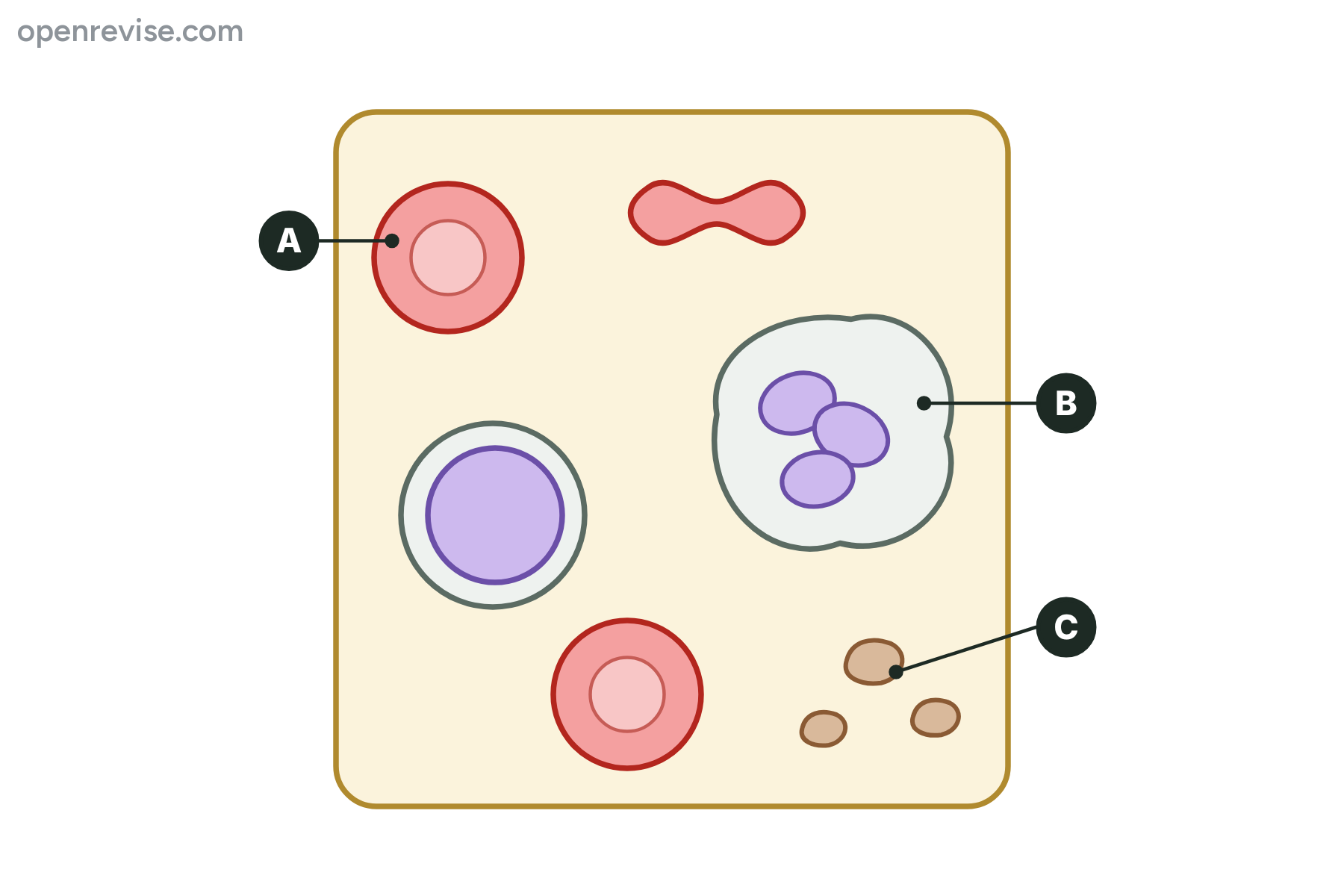
The diagram shows some of the components of blood. Name the parts labelled A, B and C.
Show answerHide answer
Model answer
A is a red blood cell. B is a phagocyte (a white blood cell). C is a platelet.
Mark scheme
- A: red blood cell — 1 mark
- B: phagocyte (white blood cell) — 1 mark
- C: platelet — 1 mark
-
3 Describe [3 marks]
Describe how platelets help to protect the body after a cut.
Show answerHide answer
Model answer
Platelets collect at the damaged blood vessel and help the blood to clot. The clot seals the wound, so the body does not lose too much blood. The clot dries to form a scab, which stops pathogens from entering the body.
Mark scheme
- Platelets help the blood to clot at the wound — 1 mark
- This stops blood loss — 1 mark
- The scab prevents pathogens from entering — 1 mark
-
4 Explain [4 marks]
Explain how the structure of a red blood cell is related to its function.
Show answerHide answer
Model answer
A red blood cell contains haemoglobin, which combines with oxygen so that the cell can carry it. It has no nucleus, so there is more space inside for haemoglobin. It is a biconcave disc, which gives it a large surface area for its volume, so oxygen can diffuse in and out quickly. It is small and flexible, so it can squeeze through the narrow capillaries to reach the tissues.
Mark scheme
- Contains haemoglobin, which combines with oxygen — 1 mark
- No nucleus, so more room for haemoglobin — 1 mark
- Biconcave disc gives a large surface area for diffusion — 1 mark
- Small and flexible to pass through capillaries — 1 mark
-
5 Explain [4 marks]
Explain how phagocytes and lymphocytes help to protect the body against pathogens.
Show answerHide answer
Model answer
A phagocyte can change its shape to flow round a pathogen and engulf it. It then digests the pathogen with enzymes. A lymphocyte makes antibodies that are specific to a pathogen. The antibodies stick to the pathogen and help to destroy it.
Mark scheme
- Phagocytes engulf pathogens — 1 mark
- They digest the pathogen — 1 mark
- Lymphocytes produce antibodies — 1 mark
- Antibodies bind to the pathogen and help to destroy it — 1 mark
-
6 Calculate [3 marks]
A student draws a white blood cell from a photomicrograph. The cell in the drawing is 48 mm across. The actual diameter of the cell is 12 \(\mu\)m. Calculate the magnification of the drawing.
Show answerHide answer
Model answer
Convert the image size to micrometres: \(48\ \text{mm} = 48 \times 1000 = 48\,000\ \mu\text{m}\). Then magnification \(= 48\,000 \div 12 = 4000\), so the magnification is \(\times 4000\).
Mark scheme
- Convert 48 mm to 48 000 \(\mu\)m — 1 mark
- \(48\,000 \div 12\) — 1 mark
- \(\times 4000\) — 1 mark
-
7 Calculate [2 marks]
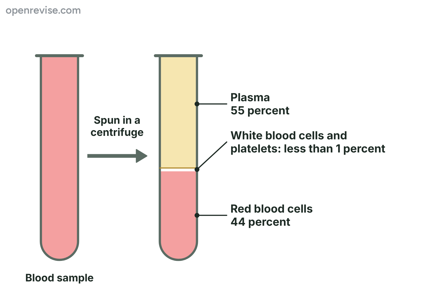
A blood sample of 600 cm\(^3\) is spun in a centrifuge and separated as shown in the diagram. Calculate the volume of plasma in the sample.
Show answerHide answer
Model answer
\(55 \div 100 \times 600 = 330\ \text{cm}^3\).
Mark scheme
- \(55 \div 100 \times 600\) — 1 mark
- 330 cm\(^3\) — 1 mark
-
8 Explain [6 marks]
Explain how the structure of the blood is related to its functions. In your answer refer to plasma, red blood cells, white blood cells and platelets.
Show answerHide answer
Model answer
Plasma is mostly water, so it dissolves and carries substances such as carbon dioxide, glucose, urea and hormones around the body. Red blood cells contain haemoglobin to carry oxygen, have no nucleus so there is more room for haemoglobin, and are biconcave discs with a large surface area for oxygen to diffuse. White blood cells have a nucleus and defend the body: phagocytes change shape to engulf and digest pathogens, and lymphocytes make antibodies to destroy them. Platelets are cell fragments that help the blood to clot, which seals wounds and stops pathogens getting in.
Mark scheme
- Plasma is mostly water, a solvent that carries dissolved substances — 1 mark
- Red blood cells contain haemoglobin to carry oxygen — 1 mark
- No nucleus and a biconcave shape suit the red cell for carrying oxygen — 1 mark
- Phagocytes change shape to engulf pathogens — 1 mark
- Lymphocytes make antibodies — 1 mark
- Platelets help blood to clot, sealing wounds and keeping pathogens out — 1 mark
Quick check
-
1
Which part of the blood carries most of the oxygen?
Show answerHide answer
C: Red blood cells
Red blood cells contain haemoglobin, which combines with oxygen.
-
2
What is haemoglobin?
Show answerHide answer
A: A red protein containing iron that combines with oxygen
Haemoglobin is a red protein that contains iron and combines with oxygen in the lungs.
-
3
Why does a red blood cell have no nucleus?
Show answerHide answer
D: So there is more room for haemoglobin
Without a nucleus there is more room for haemoglobin, so the cell carries more oxygen.
-
4
What is the job of a phagocyte?
Show answerHide answer
B: To engulf and digest pathogens
Phagocytes engulf pathogens and digest them.
-
5
Which blood component is a fragment of a cell and has no nucleus?
Show answerHide answer
A: Platelets
Platelets are fragments of cells. They have no nucleus and help the blood to clot.
-
6
Which of these is not carried in the plasma?
Show answerHide answer
D: Most of the oxygen
Oxygen is carried mainly by haemoglobin inside the red blood cells, while the other three are carried dissolved in the plasma.
-
7
A drawing of a cell is 30 mm across and the cell is 10 \(\mu\)m across in real life. What is the magnification?
Show answerHide answer
C: \(\times 3000\)
Convert 30 mm to 30 000 \(\mu\)m, then \(30\,000 \div 10 = 3000\).
-
8
Which cell makes antibodies?
Show answerHide answer
B: Lymphocyte
Lymphocytes produce antibodies that are specific to a pathogen.
Arteries, Veins and Capillaries
Just this lesson-
1 State [1 mark]
Name the type of blood vessel that carries blood away from the heart at high pressure.
Show answerHide answer
Model answer
An artery.
Mark scheme
- Artery — 1 mark
-
2 Name [3 marks]
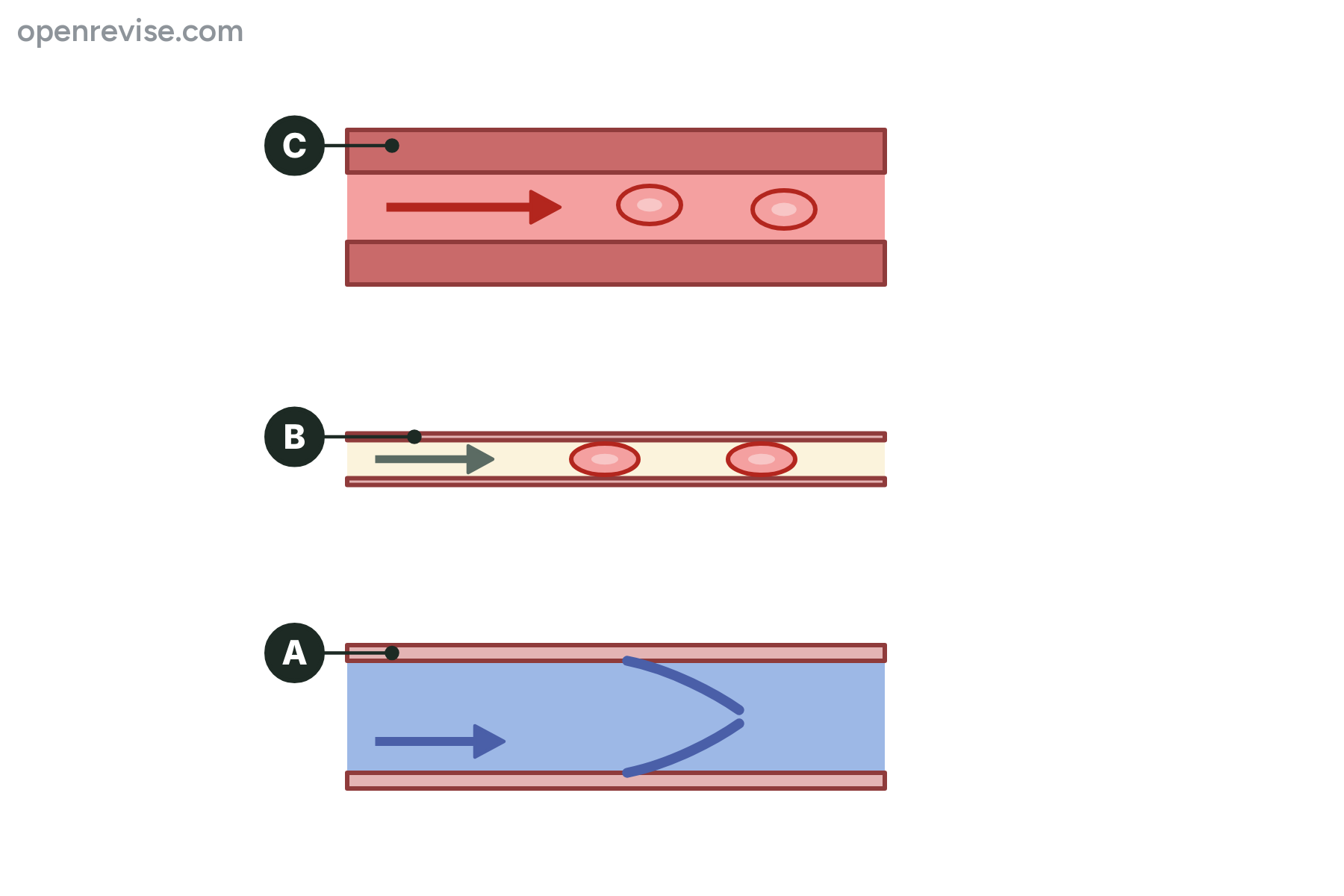
The diagram shows three types of blood vessel. Name the blood vessels labelled A, B and C.
Show answerHide answer
Model answer
A is a vein. B is a capillary. C is an artery.
Mark scheme
- A: vein — 1 mark
- B: capillary — 1 mark
- C: artery — 1 mark
-
3 Explain [2 marks]
Explain why veins have valves.
Show answerHide answer
Model answer
The blood in veins is at a low pressure, so it could flow backwards. The valves close if the blood tries to flow back, so the blood only flows towards the heart.
Mark scheme
- The pressure is low and the blood could flow backwards — 1 mark
- Valves prevent backflow, so blood flows towards the heart — 1 mark
-
4 Explain [3 marks]
Explain how the structure of a capillary is adapted for the exchange of substances between the blood and the cells.
Show answerHide answer
Model answer
The wall of a capillary is only one cell thick, so the diffusion distance is very short. Capillaries are very narrow, so red blood cells pass through in single file, close to the wall. They form a dense network, which gives a large surface area, and the blood flows slowly through them, which gives more time for exchange.
Mark scheme
- Wall one cell thick, so a short diffusion distance — 1 mark
- Very narrow, so red blood cells are close to the wall — 1 mark
- Large network gives a large surface area (and slow flow gives time for exchange) — 1 mark
-
5 Explain [4 marks]
Explain how the structure of an artery is related to its function.
Show answerHide answer
Model answer
An artery carries blood away from the heart at high pressure. It has a thick wall containing muscle, which can withstand the pressure without bursting. The wall also has elastic fibres, which stretch and recoil to smooth the flow of the blood and keep the pressure up. The lumen is narrow, which also helps to keep the pressure high.
Mark scheme
- Carries blood at high pressure — 1 mark
- Thick muscular wall withstands the pressure — 1 mark
- Elastic fibres stretch and recoil, which keeps the pressure up and smooths the flow — 1 mark
- Narrow lumen helps to maintain the pressure — 1 mark
-
6 Calculate [3 marks]
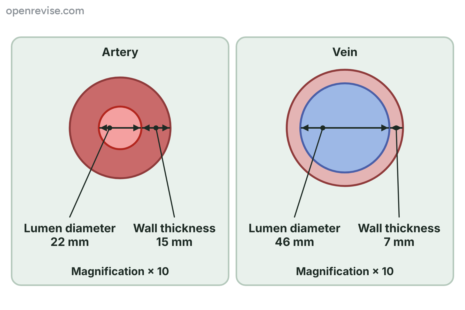
The diagram shows an artery and a vein, both magnified \(\times 10\). Calculate the actual diameter of the lumen of the vein, in mm, and the number of times wider the lumen of the vein is than the lumen of the artery. Give the second answer to 2 significant figures.
Show answerHide answer
Model answer
The actual diameter of the vein's lumen is \(46 \div 10 = 4.6\ \text{mm}\). The vein's lumen is \(46 \div 22 = 2.09\), which is 2.1 times wider than the artery's.
Mark scheme
- \(46 \div 10\) — 1 mark
- 4.6 mm — 1 mark
- 2.1 times (from \(46 \div 22\)) — 1 mark
-
7 Calculate [3 marks]
In a vein in the leg, 360 cm\(^3\) of blood flows past a point in 2 minutes. In an artery in the same leg, 480 cm\(^3\) flows past a point in 90 seconds. Calculate the rate of blood flow in cm\(^3\) per minute in each vessel.
Show answerHide answer
Model answer
Vein: \(360 \div 2 = 180\ \text{cm}^3\) per minute. Artery: \(480 \div 90 = 5.33\ \text{cm}^3\) per second, and \(5.33 \times 60 = 320\ \text{cm}^3\) per minute.
Mark scheme
- Vein: 180 cm\(^3\) per minute — 1 mark
- Artery: converts 90 seconds to 1.5 minutes, or the rate per second to per minute — 1 mark
- Artery: 320 cm\(^3\) per minute — 1 mark
-
8 Explain [6 marks]
Explain how the structure of arteries, veins and capillaries is related to the function of each type of blood vessel.
Show answerHide answer
Model answer
Arteries carry blood away from the heart at high pressure, so they have a thick wall of muscle and elastic fibres to withstand the pressure and keep it up, and a narrow lumen. Veins carry blood back to the heart at low pressure, so they have a thin wall and a wide lumen, which lets the blood flow easily, and valves that stop the blood flowing backwards. Capillaries exchange substances between the blood and the cells, so their walls are one cell thick to give a short diffusion distance, and they are very narrow and form dense networks, which gives a large surface area for exchange.
Mark scheme
- Arteries: high pressure, so a thick muscular wall — 1 mark
- Arteries: elastic fibres and narrow lumen to maintain the pressure — 1 mark
- Veins: low pressure, so a thin wall and a wide lumen — 1 mark
- Veins: valves stop backflow — 1 mark
- Capillaries: wall one cell thick, so a short diffusion distance — 1 mark
- Capillaries: narrow and in dense networks, so large surface area for exchange — 1 mark
Quick check
-
1
Which blood vessels carry blood away from the heart?
Show answerHide answer
B: Arteries
Arteries carry blood away from the heart at high pressure.
-
2
Why does the wall of an artery contain elastic fibres?
Show answerHide answer
D: To stretch and recoil with each heartbeat
The fibres stretch as each surge of blood arrives and recoil afterwards, which smooths the flow and keeps the pressure up.
-
3
What is the main function of the valves in veins?
Show answerHide answer
A: To stop the blood flowing backwards
Valves close if the blood tries to flow backwards, so it can only flow towards the heart.
-
4
How thick is the wall of a capillary?
Show answerHide answer
C: One cell thick
A capillary wall is one cell thick, which gives a short diffusion distance.
-
5
Which statement about the pulmonary vein is correct?
Show answerHide answer
B: It carries oxygenated blood to the heart
The pulmonary vein carries oxygenated blood from the lungs to the heart. It is a vein because it carries blood to the heart.
-
6
An image of a vessel is 60 mm across and the magnification is \(\times 20\). What is the actual diameter?
Show answerHide answer
A: 3 mm
Actual size equals image size divided by magnification, so \(60 \div 20 = 3\ \text{mm}\).
-
7
Which feature is found in a vein but not in an artery?
Show answerHide answer
D: Valves
Veins have valves to stop backflow. Arteries carry blood at high pressure, so they do not need them.
-
8
In 60 seconds, 240 cm\(^3\) of blood flows through a vessel. What is the rate of blood flow?
Show answerHide answer
C: 240 cm\(^3\) per minute
Rate equals volume divided by time, so \(240 \div 1 = 240\ \text{cm}^3\) per minute, which is 4 cm\(^3\) per second.
The Heart and Double Circulation
Just this lesson-
1 State [1 mark]
Name the chamber of the heart that has the thickest wall.
Show answerHide answer
Model answer
The left ventricle.
Mark scheme
- Left ventricle — 1 mark
-
2 Name [3 marks]
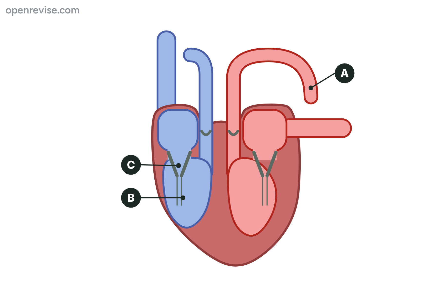
The diagram shows a section through the human heart. Name the parts labelled A, B and C.
Show answerHide answer
Model answer
A is the aorta. B is the right ventricle. C is an atrioventricular valve.
Mark scheme
- A: aorta — 1 mark
- B: right ventricle — 1 mark
- C: atrioventricular valve (valve) — 1 mark
-
3 Name [3 marks]
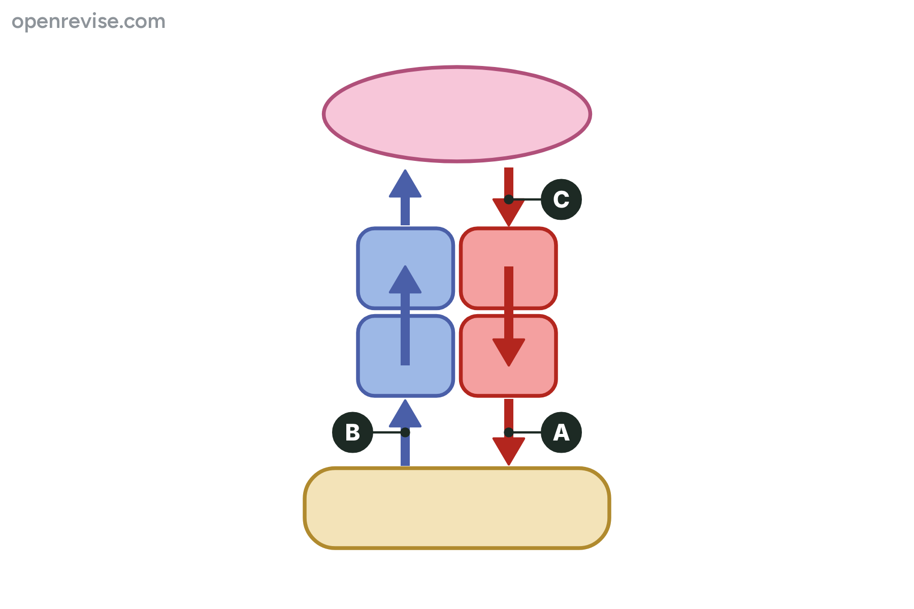
The diagram shows the circulation of the blood. Name the blood vessels labelled A, B and C.
Show answerHide answer
Model answer
A is the aorta. B is the vena cava. C is the pulmonary vein.
Mark scheme
- A: aorta — 1 mark
- B: vena cava — 1 mark
- C: pulmonary vein — 1 mark
-
4 Describe [5 marks]
Describe the route taken by blood from the vena cava to the aorta, through the heart and the lungs.
Show answerHide answer
Model answer
The blood enters the right atrium from the vena cava. It passes into the right ventricle and is pumped out through the pulmonary artery to the lungs. It returns from the lungs in the pulmonary vein to the left atrium. It passes into the left ventricle and is pumped out into the aorta.
Mark scheme
- Vena cava to the right atrium — 1 mark
- Right atrium to the right ventricle — 1 mark
- Right ventricle to the lungs through the pulmonary artery — 1 mark
- Lungs to the left atrium through the pulmonary vein — 1 mark
- Left atrium to the left ventricle, then out through the aorta — 1 mark
-
5 Explain [3 marks]
Explain why the wall of the left ventricle is thicker than the wall of the right ventricle.
Show answerHide answer
Model answer
The left ventricle pumps blood to the whole body, which is a long way, so it has to produce a high pressure. A thick muscular wall contracts with more force. The right ventricle only pumps blood to the lungs, which are close, so a lower pressure is enough and a thinner wall will do.
Mark scheme
- The left ventricle pumps blood all round the body — 1 mark
- A thick muscular wall produces a higher pressure — 1 mark
- The right ventricle only pumps blood to the lungs, so needs less pressure — 1 mark
-
6 Explain [3 marks]
Explain the function of the valves in the heart.
Show answerHide answer
Model answer
The valves make sure that the blood flows in one direction only. The atrioventricular valves close when the ventricles contract, so blood cannot flow back into the atria. The semilunar valves close when the ventricles relax, so blood cannot flow back from the arteries into the ventricles.
Mark scheme
- Valves keep the blood flowing in one direction — 1 mark
- Atrioventricular valves prevent backflow into the atria — 1 mark
- Semilunar valves prevent backflow into the ventricles — 1 mark
-
7 Explain [4 marks]
Humans have a double circulation. Explain why this is an advantage.
Show answerHide answer
Model answer
Blood passes through the heart twice on each trip round the body. After it has been through the narrow capillaries of the lungs its pressure is low, so it returns to the heart to be pumped again at a high pressure. This means that oxygenated blood reaches the body quickly. The septum keeps oxygenated and deoxygenated blood separate, so the blood leaving for the body carries as much oxygen as possible. The lungs receive blood at a lower pressure, which protects their delicate capillaries.
Mark scheme
- The blood goes through the heart twice (lungs and then body) — 1 mark
- The pressure is raised again after the lungs — 1 mark
- Oxygenated blood reaches the body faster — 1 mark
- Oxygenated and deoxygenated blood are kept separate — 1 mark
-
8 Explain [6 marks]
Explain how the structure of the heart and the circulatory system is related to their function.
Show answerHide answer
Model answer
The heart has four chambers. The atria have thin walls because they only push blood into the ventricles, and the ventricles have thicker walls because they pump blood out of the heart. The left ventricle has the thickest wall because it pumps blood at high pressure round the whole body, while the right ventricle pumps blood only to the lungs. The septum keeps oxygenated and deoxygenated blood apart. Valves between the atria and ventricles, and in the arteries, stop blood flowing backwards. Blood goes to the lungs through the pulmonary artery and returns in the pulmonary vein, and leaves for the body through the aorta and returns in the vena cava. This double circulation lets the pressure be raised again after the lungs.
Mark scheme
- Thin atria and thicker ventricles, linked to function — 1 mark
- Left ventricle thicker than right, linked to the pressure needed — 1 mark
- Septum separates oxygenated and deoxygenated blood — 1 mark
- Valves stop backflow — 1 mark
- Correct route using the main vessels — 1 mark
- Double circulation restores the pressure — 1 mark
Quick check
-
1
Which vessel carries oxygenated blood from the heart to the body?
Show answerHide answer
D: Aorta
The aorta carries oxygenated blood from the left ventricle to the body.
-
2
Which vessel brings blood from the lungs to the heart?
Show answerHide answer
A: Pulmonary vein
The pulmonary vein carries oxygenated blood from the lungs to the left atrium.
-
3
What is the main function of the septum?
Show answerHide answer
C: To keep the two sides of the heart separate
The septum separates the right and left sides of the heart so oxygenated and deoxygenated blood do not mix.
-
4
Why do the atria have thinner walls than the ventricles?
Show answerHide answer
B: They only push blood into the ventricles
The atria only push blood a short way into the ventricles, but the ventricles must pump it out of the heart.
-
5
What do the semilunar valves prevent?
Show answerHide answer
D: Blood flowing back into the ventricles from the arteries
They close when the ventricles relax, so blood cannot flow back from the arteries into the ventricles.
-
6
Where does blood pick up oxygen in the double circulation?
Show answerHide answer
C: In the lungs
Blood picks up oxygen in the capillaries of the lungs, between the pulmonary artery and the pulmonary vein.
-
7
Why is the circulatory system of a human called a double circulation?
Show answerHide answer
A: Blood passes through the heart twice on each trip
The blood passes through the heart twice on each complete trip, once going to the lungs and once going to the body.
-
8
A baby is born with a hole in the septum. Which problem is this most likely to cause?
Show answerHide answer
B: The blood sent to the body carries less oxygen
Oxygenated and deoxygenated blood can mix, so the blood sent to the body carries less oxygen.
Heart Rate, Stroke Volume and Cardiac Output
Just this lesson-
1 State [1 mark]
Write down the equation that links cardiac output, stroke volume and heart rate.
Show answerHide answer
Model answer
Cardiac output = stroke volume \(\times\) heart rate.
Mark scheme
- Cardiac output = stroke volume \(\times\) heart rate — 1 mark
-
2 Calculate [2 marks]
A person has a heart rate of 75 beats per minute and a stroke volume of 68 cm\(^3\). Calculate the cardiac output in cm\(^3\) per minute.
Show answerHide answer
Model answer
Cardiac output \(= 68 \times 75 = 5100\ \text{cm}^3\) per minute.
Mark scheme
- \(68 \times 75\) — 1 mark
- 5100 cm\(^3\) per minute — 1 mark
-
3 Calculate [3 marks]
A person has a cardiac output of 4.8 dm\(^3\) per minute and a heart rate of 80 beats per minute. Calculate the stroke volume of the person in cm\(^3\).
Show answerHide answer
Model answer
Convert the cardiac output: \(4.8 \times 1000 = 4800\ \text{cm}^3\) per minute. Stroke volume \(= 4800 \div 80 = 60\ \text{cm}^3\).
Mark scheme
- Converts 4.8 dm\(^3\) to 4800 cm\(^3\) — 1 mark
- \(4800 \div 80\) — 1 mark
- 60 cm\(^3\) — 1 mark
-
4 Calculate [3 marks]
A student counts 18 pulse beats in 15 seconds. Her stroke volume is 65 cm\(^3\). Calculate her heart rate in beats per minute and her cardiac output in cm\(^3\) per minute.
Show answerHide answer
Model answer
Heart rate \(= 18 \times 4 = 72\) beats per minute. Cardiac output \(= 65 \times 72 = 4680\ \text{cm}^3\) per minute.
Mark scheme
- \(18 \times 4\) — 1 mark
- Heart rate 72 beats per minute — 1 mark
- Cardiac output 4680 cm\(^3\) per minute — 1 mark
-
5 Describe [4 marks]
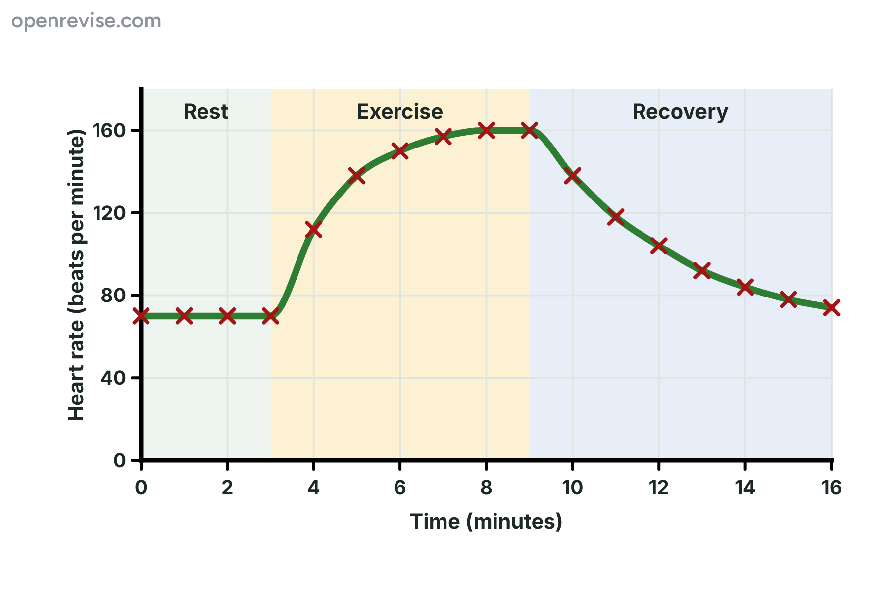
The graph shows the heart rate of a person before, during and after exercise. Describe the change in heart rate during the exercise period, and calculate the percentage increase in heart rate from the resting rate to the highest rate.
Show answerHide answer
Model answer
During exercise the heart rate rises quickly from 70 to about 138 beats per minute in the first two minutes, then more slowly, and levels off at 160 beats per minute. The increase is \(160 - 70 = 90\) beats per minute, so the percentage increase is \(90 \div 70 \times 100 = 128.6\), which is 129 percent.
Mark scheme
- Rises quickly at first, then more slowly — 1 mark
- Levels off at 160 beats per minute — 1 mark
- \(90 \div 70 \times 100\) — 1 mark
- 129 percent — 1 mark
-
6 Explain [3 marks]
Explain why the heart rate and the stroke volume of a person increase during exercise.
Show answerHide answer
Model answer
The muscles respire faster, so they need more oxygen and glucose and produce more carbon dioxide. The nervous system and adrenaline make the heart beat faster, so the heart rate rises. The heart muscle also contracts more strongly, so the stroke volume rises. Both changes increase the cardiac output, so more blood reaches the muscles.
Mark scheme
- Muscles need more oxygen and glucose, and have more carbon dioxide to remove — 1 mark
- Heart rate rises so the heart beats faster — 1 mark
- Stroke volume rises because the heart muscle contracts more strongly — 1 mark
-
7 Calculate [4 marks]
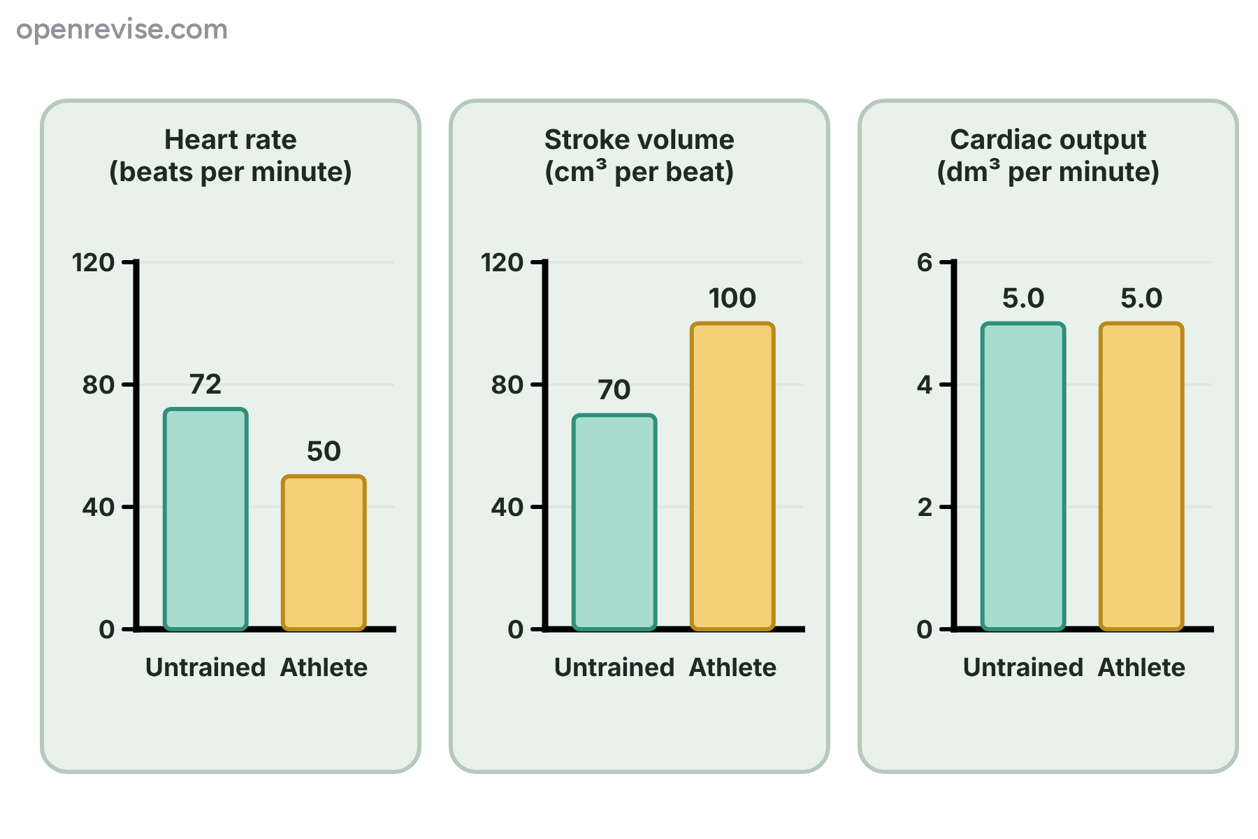
The charts show the heart rate, stroke volume and cardiac output of an untrained person and an athlete at rest. Calculate the cardiac output of the athlete in cm\(^3\) per minute, and explain why the athlete has a lower heart rate than the untrained person although the cardiac output is the same.
Show answerHide answer
Model answer
Cardiac output of the athlete \(= 100 \times 50 = 5000\ \text{cm}^3\) per minute. The athlete has a larger stroke volume, because training has made the heart muscle stronger. Each beat pumps out more blood, so fewer beats per minute are needed to give the same cardiac output.
Mark scheme
- \(100 \times 50\) — 1 mark
- 5000 cm\(^3\) per minute — 1 mark
- The athlete has a larger stroke volume — 1 mark
- So fewer beats are needed to pump the same volume of blood per minute — 1 mark
-
8 Evaluate [6 marks]
Source: A volunteer trained on a bicycle for six months. Before training: at rest the heart rate was 75 beats per minute and the stroke volume was 64 cm\(^3\). During hard exercise the heart rate was 170 beats per minute and the stroke volume was 105 cm\(^3\). After training: at rest the heart rate was 60 beats per minute and the stroke volume was 80 cm\(^3\). During hard exercise the heart rate was 172 beats per minute and the stroke volume was 130 cm\(^3\). Use the data to evaluate the effect of six months of training on the heart of the volunteer. Include calculations of cardiac output in your answer.
Show answerHide answer
Model answer
At rest, before training the cardiac output was \(64 \times 75 = 4800\ \text{cm}^3\) per minute and after training it was \(80 \times 60 = 4800\ \text{cm}^3\) per minute, so it was unchanged. The resting heart rate fell because the stroke volume rose, so fewer beats were needed. During hard exercise the cardiac output rose from \(105 \times 170 = 17\,850\ \text{cm}^3\) per minute to \(130 \times 172 = 22\,360\ \text{cm}^3\) per minute, an increase of 4510 cm\(^3\) per minute, which is about 25 percent. Training has made the heart muscle stronger, so the stroke volume is larger. This means that more oxygen and glucose are delivered to the muscles during exercise, so the volunteer can exercise harder for longer. The effect on the heart is beneficial.
Mark scheme
- Resting cardiac output is 4800 before and after training — 1 mark
- Resting heart rate falls because the stroke volume increases — 1 mark
- Exercise cardiac output rises from 17 850 to 22 360 cm\(^3\) per minute — 1 mark
- An increase of about 25 percent — 1 mark
- Training strengthens the heart muscle, giving a larger stroke volume — 1 mark
- More oxygen is delivered to the muscles, so performance improves (a conclusion that the training is beneficial) — 1 mark
Quick check
-
1
What is the unit of heart rate?
Show answerHide answer
C: Beats per minute
Heart rate is the number of beats in one minute, so it is measured in beats per minute.
-
2
Which equation is correct?
Show answerHide answer
A: Cardiac output = stroke volume \(\times\) heart rate
Cardiac output is the stroke volume multiplied by the heart rate.
-
3
A person has a stroke volume of 70 cm\(^3\) and a heart rate of 60 beats per minute. What is the cardiac output?
Show answerHide answer
D: 4200 cm\(^3\) per minute
\(70 \times 60 = 4200\ \text{cm}^3\) per minute.
-
4
A person's cardiac output is 6000 cm\(^3\) per minute and their heart rate is 75 beats per minute. What is the stroke volume?
Show answerHide answer
B: 80 cm\(^3\)
Stroke volume is cardiac output divided by heart rate, so \(6000 \div 75 = 80\ \text{cm}^3\).
-
5
A pulse is counted for 30 seconds and there are 36 beats. What is the heart rate?
Show answerHide answer
C: 72 beats per minute
30 seconds is half a minute, so double the count: \(36 \times 2 = 72\) beats per minute.
-
6
What happens to the cardiac output during exercise?
Show answerHide answer
A: It increases
Both the heart rate and the stroke volume increase, so the cardiac output increases.
-
7
Why does a trained athlete have a lower resting heart rate than an untrained person?
Show answerHide answer
D: The athlete has a larger stroke volume
Training gives a stronger heart with a larger stroke volume, so fewer beats are needed to give the same cardiac output.
-
8
What does the recovery time measure?
Show answerHide answer
B: The time for the heart rate to return to normal after exercise
The recovery time is how long the heart rate takes to return to its resting value after exercise stops.